1VDX | pdb_00001vdx

Crystal Structure of a Pyrococcus horikoshii protein with similarities to 2'5' RNA-ligase


Experimental Data Snapshot

  • Method: X-RAY DIFFRACTION
  • Resolution: 2.40 Å
  • R-Value Free: 
    0.274 (Depositor), 0.276 (DCC) 
  • R-Value Work: 
    0.225 (Depositor), 0.226 (DCC) 
  • R-Value Observed: 
    0.225 (Depositor) 

Starting Model: experimental
View more details

wwPDB Validation 3D Report Full Report

Validation slider image for 1VDX

This is version 1.3 of the entry. See complete history

Literature

Structure of a putative 2'-5' RNA ligase from Pyrococcus horikoshii.

Rehse, P.H.Tahirov, T.H.

(2005) Acta Crystallogr D Biol Crystallogr 61: 1207-1212

  • DOI: https://doi.org/10.1107/S0907444905017841
  • Primary Citation Related Structures: 
    1VDX

  • PubMed Abstract: 

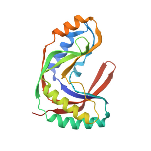
    Cyclic phosphodiesterase and 2'-5' RNA ligase are members of a superfamily of proteins which share structural similarities even though their homology may be very low. A putative 2'-5' RNA ligase from Pyrococcus horikoshii has been crystallized and its X-ray crystallographic structure determined to 2.4 A. The protein crystallized in the orthorhombic space group P2(1)2(1)2(1), with unit-cell parameters a = 44.07, b = 45.47, c = 93.17 A and one protein monomer in the asymmetric unit. The molecular-replacement probe was a 2'-5' RNA ligase from Thermus thermophilus which shares 30% sequence identity. The P. horikoshii RNA ligase has some structural features that have more in common with a cyclic phosphodiesterase from Arabidopsis thaliana with which it has no significant homology, yet an examination of the electrostatic surface potential clearly defines its relationship to the T. thermophilus RNA ligase. However, the size of the active-site cleft is smaller and less positively charged than that of the T. thermophilus homologue, suggesting that the actual substrate may be smaller than that previously postulated for the latter.


  • Organizational Affiliation
    • RIKEN Harima Institute, 1-1-1 Kouto, Mikazuki-cho, Sayo-gun, Hyogo 679-5148, Japan.

Macromolecule Content 

  • Total Structure Weight: 21.17 kDa 
  • Atom Count: 1,530 
  • Modeled Residue Count: 184 
  • Deposited Residue Count: 184 
  • Unique protein chains: 1

Macromolecules

Find similar proteins by:|  3D Structure
Entity ID: 1
MoleculeChains  Sequence LengthOrganismDetailsImage
Hypothetical protein PH0099184Pyrococcus horikoshiiMutation(s): 0 
EC: 6.5.1.3 (PDB Primary Data), 3.1.4.58 (UniProt)
UniProt
Find proteins for O57823 (Pyrococcus horikoshii (strain ATCC 700860 / DSM 12428 / JCM 9974 / NBRC 100139 / OT-3))
Explore O57823 
Go to UniProtKB:  O57823
Entity Groups
Sequence Clusters30% Identity50% Identity70% Identity90% Identity95% Identity100% Identity
UniProt GroupO57823
Sequence Annotations
Expand
Reference Sequence

Small Molecules

Ligands 1 Unique
IDChains Name / Formula / InChI Key2D Diagram3D Interactions
CL

Query on CL


Download:Ideal Coordinates CCD File
B [auth A]CHLORIDE ION
Cl
VEXZGXHMUGYJMC-UHFFFAOYSA-M

Experimental Data & Validation

Experimental Data

  • Method: X-RAY DIFFRACTION
  • Resolution: 2.40 Å
  • R-Value Free:  0.274 (Depositor), 0.276 (DCC) 
  • R-Value Work:  0.225 (Depositor), 0.226 (DCC) 
  • R-Value Observed: 0.225 (Depositor) 
Space Group: P 21 21 21
Unit Cell:
Length ( Å )Angle ( ˚ )
a = 44.068α = 90
b = 45.471β = 90
c = 93.171γ = 90
Software Package:
Software NamePurpose
CNSrefinement
HKL-2000data reduction
SCALEPACKdata scaling
MOLREPphasing

Structure Validation

View Full Validation Report


Entry History 

Deposition Data

Revision History  (Full details and data files)

  • Version 1.0: 2005-04-12
    Type: Initial release
  • Version 1.1: 2008-04-27
    Changes: Version format compliance
  • Version 1.2: 2011-07-13
    Changes: Version format compliance
  • Version 1.3: 2023-10-25
    Changes: Data collection, Database references, Refinement description