1V9P | pdb_00001v9p

Crystal Structure Of Nad+-Dependent DNA Ligase


Experimental Data Snapshot

  • Method: X-RAY DIFFRACTION
  • Resolution: 2.90 Å
  • R-Value Free: 
    0.293 (Depositor), 0.303 (DCC) 
  • R-Value Work: 
    0.232 (Depositor), 0.244 (DCC) 
  • R-Value Observed: 
    0.232 (Depositor) 

wwPDB Validation 3D Report Full Report

Validation slider image for 1V9P

Ligand Structure Quality Assessment 


This is version 1.3 of the entry. See complete history

Literature

Crystal structure of NAD(+)-dependent DNA ligase: modular architecture and functional implications.

Lee, J.Y.Chang, C.Song, H.K.Moon, J.Yang, J.K.Kim, H.K.Kwon, S.T.Suh, S.W.

(2000) EMBO J 19: 1119-1129

  • DOI: https://doi.org/10.1093/emboj/19.5.1119
  • Primary Citation Related Structures: 
    1DGS, 1V9P

  • PubMed Abstract: 

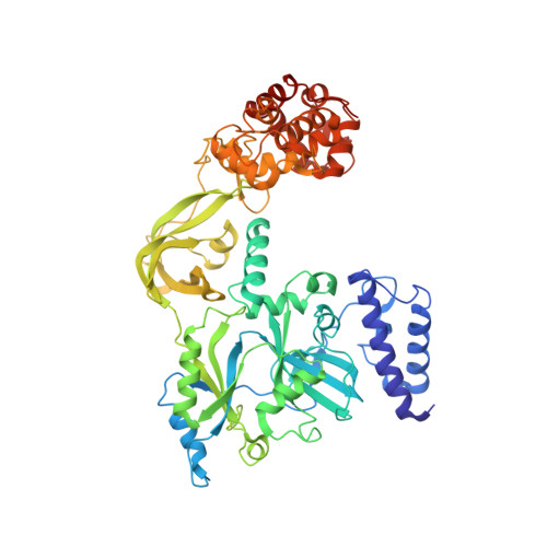
    DNA ligases catalyze the crucial step of joining the breaks in duplex DNA during DNA replication, repair and recombination, utilizing either ATP or NAD(+) as a cofactor. Despite the difference in cofactor specificity and limited overall sequence similarity, the two classes of DNA ligase share basically the same catalytic mechanism. In this study, the crystal structure of an NAD(+)-dependent DNA ligase from Thermus filiformis, a 667 residue multidomain protein, has been determined by the multiwavelength anomalous diffraction (MAD) method. It reveals highly modular architecture and a unique circular arrangement of its four distinct domains. It also provides clues for protein flexibility and DNA-binding sites. A model for the multidomain ligase action involving large conformational changes is proposed.


  • Organizational Affiliation
    • Center for Molecular Catalysis, Department of Chemistry, College of Natural Sciences, Seoul National University, Seoul 151-742.

Macromolecule Content 

  • Total Structure Weight: 135.47 kDa 
  • Atom Count: 9,768 
  • Modeled Residue Count: 1,168 
  • Deposited Residue Count: 1,168 
  • Unique protein chains: 1

Macromolecules

Find similar proteins by:|  3D Structure
Entity ID: 1
MoleculeChains  Sequence LengthOrganismDetailsImage
DNA ligase
A, B
584Thermus filiformisMutation(s): 0 
Gene Names: LIGA
EC: 6.5.1.2
UniProt
Find proteins for Q9ZHI0 (Thermus filiformis)
Explore Q9ZHI0 
Go to UniProtKB:  Q9ZHI0
Entity Groups
Sequence Clusters30% Identity50% Identity70% Identity90% Identity95% Identity100% Identity
UniProt GroupQ9ZHI0
Sequence Annotations
Expand
Reference Sequence

Experimental Data & Validation

Experimental Data

  • Method: X-RAY DIFFRACTION
  • Resolution: 2.90 Å
  • R-Value Free:  0.293 (Depositor), 0.303 (DCC) 
  • R-Value Work:  0.232 (Depositor), 0.244 (DCC) 
  • R-Value Observed: 0.232 (Depositor) 
Space Group: P 1 21 1
Unit Cell:
Length ( Å )Angle ( ˚ )
a = 89.21α = 90
b = 117.33β = 115.09
c = 97.48γ = 90
Software Package:
Software NamePurpose
CNSrefinement
DENZOdata reduction
SCALEPACKdata scaling
SOLVEphasing

Structure Validation

View Full Validation Report


Ligand Structure Quality Assessment 


Entry History 

Deposition Data

Revision History  (Full details and data files)

  • Version 1.0: 2004-03-30
    Type: Initial release
  • Version 1.1: 2008-04-27
    Changes: Version format compliance
  • Version 1.2: 2011-07-13
    Changes: Version format compliance
  • Version 1.3: 2023-12-27
    Changes: Data collection, Database references, Derived calculations