1V4A | pdb_00001v4a

Structure of the N-terminal Domain of Escherichia coli Glutamine Synthetase adenylyltransferase


Experimental Data Snapshot

  • Method: X-RAY DIFFRACTION
  • Resolution: 2.00 Å
  • R-Value Free: 
    0.269 (Depositor), 0.277 (DCC) 
  • R-Value Work: 
    0.228 (Depositor), 0.239 (DCC) 
  • R-Value Observed: 
    0.230 (Depositor) 

wwPDB Validation 3D Report Full Report

Validation slider image for 1V4A

This is version 1.4 of the entry. See complete history

Literature

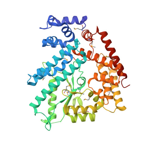
Structure of the n-terminal domain of Escherichia coli glutamine synthetase adenylyltransferase

Xu, Y.Zhang, R.Joachimiak, A.Carr, P.D.Huber, T.Vasudevan, S.G.Ollis, D.L.

(2004) Structure 12: 861-869

  • DOI: https://doi.org/10.1016/j.str.2004.02.029
  • Primary Citation Related Structures: 
    1V4A

  • PubMed Abstract: 

    We report the crystal structure of the N-terminal domain of Escherichia coli adenylyltransferase that catalyzes the reversible nucleotidylation of glutamine synthetase (GS), a key enzyme in nitrogen assimilation. This domain (AT-N440) catalyzes the deadenylylation and subsequent activation of GS. The structure has been divided into three subdomains, two of which bear some similarity to kanamycin nucleotidyltransferase (KNT). However, the orientation of the two domains in AT-N440 differs from that in KNT. The active site of AT-N440 has been identified on the basis of structural comparisons with KNT, DNA polymerase beta, and polyadenylate polymerase. AT-N440 has a cluster of metal binding residues that are conserved in polbeta-like nucleotidyl transferases. The location of residues conserved in all ATase sequences was found to cluster around the active site. Many of these residues are very likely to play a role in catalysis, substrate binding, or effector binding.


  • Organizational Affiliation
    • Department of Biochemistry and Molecular Biology, James Cook University, Townsville, Queensland 4811, Australia. yibin.xu@jcu.edu.au

Macromolecule Content 

  • Total Structure Weight: 51.04 kDa 
  • Atom Count: 3,541 
  • Modeled Residue Count: 429 
  • Deposited Residue Count: 440 
  • Unique protein chains: 1

Macromolecules

Find similar proteins by:|  3D Structure
Entity ID: 1
MoleculeChains  Sequence LengthOrganismDetailsImage
Glutamate-ammonia-ligase adenylyltransferase440Escherichia coliMutation(s): 13 
EC: 2.7.7.42 (PDB Primary Data), 2.7.7.89 (UniProt)
UniProt
Find proteins for P30870 (Escherichia coli (strain K12))
Explore P30870 
Go to UniProtKB:  P30870
Entity Groups
Sequence Clusters30% Identity50% Identity70% Identity90% Identity95% Identity100% Identity
UniProt GroupP30870
Sequence Annotations
Expand
Reference Sequence

Small Molecules

Modified Residues  1 Unique
IDChains TypeFormula2D DiagramParent
MSE
Query on MSE
A
L-PEPTIDE LINKINGC5 H11 N O2 SeMET

Experimental Data & Validation

Experimental Data

  • Method: X-RAY DIFFRACTION
  • Resolution: 2.00 Å
  • R-Value Free:  0.269 (Depositor), 0.277 (DCC) 
  • R-Value Work:  0.228 (Depositor), 0.239 (DCC) 
  • R-Value Observed: 0.230 (Depositor) 
Space Group: P 32 2 1
Unit Cell:
Length ( Å )Angle ( ˚ )
a = 116.836α = 90
b = 116.836β = 90
c = 67.744γ = 120
Software Package:
Software NamePurpose
REFMACrefinement
HKL-2000data reduction
SCALEPACKdata scaling
SOLVEphasing

Structure Validation

View Full Validation Report


Entry History 

Deposition Data

Revision History  (Full details and data files)

  • Version 1.0: 2004-07-27
    Type: Initial release
  • Version 1.1: 2008-04-27
    Changes: Version format compliance
  • Version 1.2: 2011-07-13
    Changes: Version format compliance
  • Version 1.3: 2023-12-27
    Changes: Data collection, Database references, Derived calculations
  • Version 1.4: 2024-11-13
    Changes: Structure summary