1V1Q | pdb_00001v1q

Crystal structure of PriB- a primosomal DNA replication protein of Escherichia coli


Experimental Data Snapshot

  • Method: X-RAY DIFFRACTION
  • Resolution: 2.10 Å
  • R-Value Free: 
    0.263 (Depositor), 0.257 (DCC) 
  • R-Value Work: 
    0.206 (Depositor), 0.202 (DCC) 
  • R-Value Observed: 
    0.206 (Depositor) 

wwPDB Validation 3D Report Full Report

Validation slider image for 1V1Q

This is version 1.4 of the entry. See complete history

Literature

Crystal Structure of Prib- a Primosomal DNA Replication Protein of Escherichia Coli

Liu, J.-H.Chang, T.-W.Huang, C.-Y.Chen, S.-U.Wu, H.-N.Chang, M.-C.Hsiao, C.-D.

(2004) J Biological Chem 279: 50465

  • DOI: https://doi.org/10.1074/jbc.M406773200
  • Primary Citation Related Structures: 
    1V1Q

  • PubMed Abstract: 

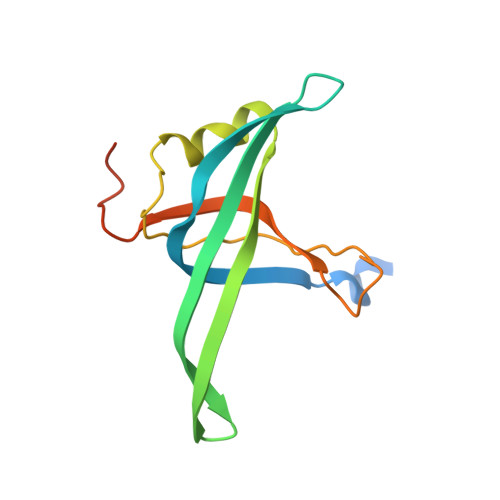
    PriB is one of the Escherichia coli varphiX-type primosome proteins that are required for assembly of the primosome, a mobile multi-enzyme complex responsible for the initiation of DNA replication. Here we report the crystal structure of the E. coli PriB at 2.1 A resolution by multi-wavelength anomalous diffraction using a mercury derivative. The polypeptide chain of PriB is structurally similar to that of single-stranded DNA-binding protein (SSB). However, the biological unit of PriB is a dimer, not a homotetramer like SSB. Electrophoretic mobility shift assays demonstrated that PriB binds single-stranded DNA and single-stranded RNA with comparable affinity. We also show that PriB binds single-stranded DNA with certain base preferences. Based on the PriB structural information and biochemical studies, we propose that the potential tetramer formation surface and several other regions of PriB may participate in protein-protein interaction during DNA replication. These findings may illuminate the role of PriB in varphiX-type primosome assembly.


  • Organizational Affiliation
    • Graduate Institute of Life Sciences, National Defense Medical Center, Taipei, Taiwan 114.

Macromolecule Content 

  • Total Structure Weight: 29.95 kDa 
  • Atom Count: 1,859 
  • Modeled Residue Count: 223 
  • Deposited Residue Count: 268 
  • Unique protein chains: 1

Macromolecules

Find similar proteins by:|  3D Structure
Entity ID: 1
MoleculeChains  Sequence LengthOrganismDetailsImage
PRIMOSOMAL REPLICATION PROTEIN N
A, B
134Escherichia coli K-12Mutation(s): 1 
UniProt
Find proteins for P07013 (Escherichia coli (strain K12))
Explore P07013 
Go to UniProtKB:  P07013
Entity Groups
Sequence Clusters30% Identity50% Identity70% Identity90% Identity95% Identity100% Identity
UniProt GroupP07013
Sequence Annotations
Expand
Reference Sequence

Experimental Data & Validation

Experimental Data

  • Method: X-RAY DIFFRACTION
  • Resolution: 2.10 Å
  • R-Value Free:  0.263 (Depositor), 0.257 (DCC) 
  • R-Value Work:  0.206 (Depositor), 0.202 (DCC) 
  • R-Value Observed: 0.206 (Depositor) 
Space Group: P 21 21 21
Unit Cell:
Length ( Å )Angle ( ˚ )
a = 39.322α = 90
b = 57.358β = 90
c = 97.019γ = 90
Software Package:
Software NamePurpose
CNSrefinement
DENZOdata reduction
SCALEPACKdata scaling
SOLVEphasing

Structure Validation

View Full Validation Report


Entry History 

Deposition Data

Revision History  (Full details and data files)

  • Version 1.0: 2004-10-25
    Type: Initial release
  • Version 1.1: 2011-05-08
    Changes: Version format compliance
  • Version 1.2: 2011-07-13
    Changes: Version format compliance
  • Version 1.3: 2023-11-08
    Changes: Data collection, Database references, Derived calculations, Other
  • Version 1.4: 2024-11-20
    Changes: Structure summary