1V04 | pdb_00001v04

serum paraoxonase by directed evolution


Experimental Data Snapshot

  • Method: X-RAY DIFFRACTION
  • Resolution: 2.20 Å
  • R-Value Free: 
    0.217 (Depositor), 0.239 (DCC) 
  • R-Value Work: 
    0.185 (Depositor), 0.234 (DCC) 
  • R-Value Observed: 
    0.186 (Depositor) 

wwPDB Validation 3D Report Full Report

Validation slider image for 1V04

This is version 1.2 of the entry. See complete history

Literature

Structure and Evolution of the Serum Paraoxonase Family of Detoxifying and Anti-Atherosclerotic Enzymes

Harel, M.Aharoni, A.Gaidukov, L.Brumshtein, B.Khersonsky, O.Meged, R.Dvir, H.Ravelli, R.B.G.Mccarthy, A.Toker, L.Silman, I.Sussman, J.L.Tawfik, D.S.

(2004) Nat Struct Mol Biol 11: 412

  • DOI: https://doi.org/10.1038/nsmb767
  • Primary Citation Related Structures: 
    1V04

  • PubMed Abstract: 

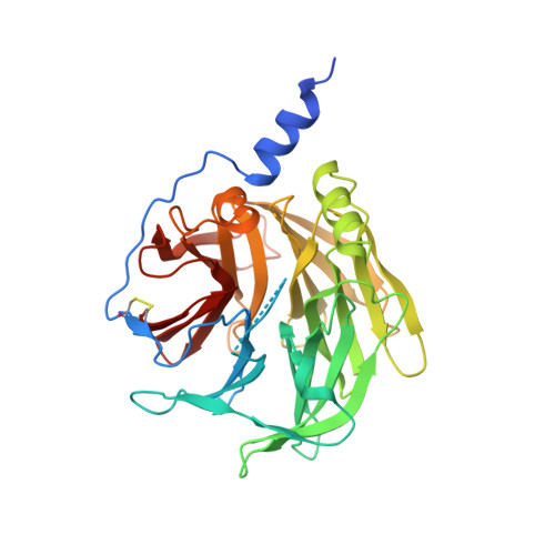
    Members of the serum paraoxonase (PON) family have been identified in mammals and other vertebrates, and in invertebrates. PONs exhibit a wide range of physiologically important hydrolytic activities, including drug metabolism and detoxification of nerve agents. PON1 and PON3 reside on high-density lipoprotein (HDL, 'good cholesterol') and are involved in the prevention of atherosclerosis. We describe the first crystal structure of a PON family member, a variant of PON1 obtained by directed evolution, at a resolution of 2.2 A. PON1 is a six-bladed beta-propeller with a unique active site lid that is also involved in HDL binding. The three-dimensional structure and directed evolution studies permit a detailed description of PON1's active site and catalytic mechanism, which are reminiscent of secreted phospholipase A2, and of the routes by which PON family members diverged toward different substrate and reaction selectivities.


  • Organizational Affiliation
    • Department of Structural Biology, The Weizmann Institute of Science, Rehovot 76 100, Israel.

Macromolecule Content 

  • Total Structure Weight: 39.78 kDa 
  • Atom Count: 2,756 
  • Modeled Residue Count: 332 
  • Deposited Residue Count: 355 
  • Unique protein chains: 1

Macromolecules

Find similar proteins by:|  3D Structure
Entity ID: 1
MoleculeChains  Sequence LengthOrganismDetailsImage
SERUM PARAOXONASE/ARYLESTERASE 1355Homo sapiensOryctolagus cuniculusMus musculusRattus rattus
This entity is chimeric
Mutation(s): 0 
EC: 3.1.1.2 (PDB Primary Data), 3.1.8.1 (PDB Primary Data), 3.1.1.81 (UniProt)
Membrane Entity: Yes 
UniProt
Find proteins for P27170 (Oryctolagus cuniculus)
Explore P27170 
Go to UniProtKB:  P27170
Entity Groups
Sequence Clusters30% Identity50% Identity70% Identity90% Identity95% Identity100% Identity
UniProt GroupP27170
Sequence Annotations
Expand
Reference Sequence

Experimental Data & Validation

Experimental Data

  • Method: X-RAY DIFFRACTION
  • Resolution: 2.20 Å
  • R-Value Free:  0.217 (Depositor), 0.239 (DCC) 
  • R-Value Work:  0.185 (Depositor), 0.234 (DCC) 
  • R-Value Observed: 0.186 (Depositor) 
Space Group: P 43 21 2
Unit Cell:
Length ( Å )Angle ( ˚ )
a = 98.44α = 90
b = 98.44β = 90
c = 139.17γ = 90
Software Package:
Software NamePurpose
REFMACrefinement
XDSdata reduction
XSCALEdata scaling
SHARPphasing

Structure Validation

View Full Validation Report


Entry History 

Revision History  (Full details and data files)

  • Version 1.0: 2004-04-23
    Type: Initial release
  • Version 1.1: 2017-03-22
    Changes: Source and taxonomy
  • Version 1.2: 2024-10-23
    Changes: Data collection, Database references, Derived calculations, Other, Structure summary