1UV6 | pdb_00001uv6

X-ray structure of acetylcholine binding protein (AChBP) in complex with carbamylcholine


Experimental Data Snapshot

  • Method: X-RAY DIFFRACTION
  • Resolution: 2.50 Å
  • R-Value Free: 
    0.265 (Depositor) 
  • R-Value Work: 
    0.227 (Depositor) 

Starting Model: experimental
View more details

wwPDB Validation 3D Report Full Report

Validation slider image for 1UV6

This is version 1.3 of the entry. See complete history

Literature

Nicotine and Carbamylcholine Binding to Nicotinic Acetylcholine Receptors as Studied in Achbp Crystal Structures

Celie, P.H.N.Van Rossum-Fikkert, S.E.Van Dijk, W.J.Brejc, K.Smit, A.B.Sixma, T.K.

(2004) Neuron 41: 907

  • DOI: https://doi.org/10.1016/s0896-6273(04)00115-1
  • Primary Citation Related Structures: 
    1UV6, 1UW6, 1UX2

  • PubMed Abstract: 

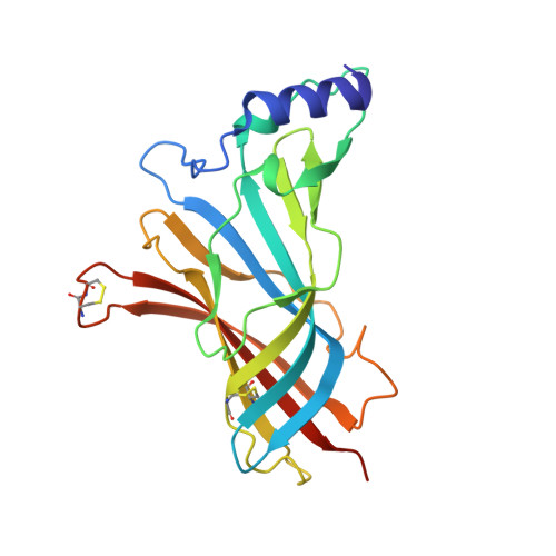
    Nicotinic acetylcholine receptors are prototypes for the pharmaceutically important family of pentameric ligand-gated ion channels. Here we present atomic resolution structures of nicotine and carbamylcholine binding to AChBP, a water-soluble homolog of the ligand binding domain of nicotinic receptors and their family members, GABAA, GABAC, 5HT3 serotonin, and glycine receptors. Ligand binding is driven by enthalpy and is accompanied by conformational changes in the ligand binding site. Residues in the binding site contract around the ligand, with the largest movement in the C loop. As expected, the binding is characterized by substantial aromatic and hydrophobic contributions, but additionally there are close contacts between protein oxygens and positively charged groups in the ligands. The higher affinity of nicotine is due to a main chain hydrogen bond with the B loop and a closer packing of the aromatic groups. These structures will be useful tools for the development of new drugs involving nicotinic acetylcholine receptor-associated diseases.


  • Organizational Affiliation
    • Division of Molecular Carcinogenesis, Netherlands Cancer Institute, Plesmanlaan 121, 1066 CX Amsterdam, The Netherlands.

Macromolecule Content 

  • Total Structure Weight: 239.07 kDa 
  • Atom Count: 16,488 
  • Modeled Residue Count: 2,050 
  • Deposited Residue Count: 2,100 
  • Unique protein chains: 1

Macromolecules

Find similar proteins by:|  3D Structure
Entity ID: 1
MoleculeChains  Sequence LengthOrganismDetailsImage
ACETYLCHOLINE-BINDING PROTEIN
A, B, C, D, E
A, B, C, D, E, F, G, H, I, J
210Lymnaea stagnalisMutation(s): 0 
UniProt
Find proteins for P58154 (Lymnaea stagnalis)
Explore P58154 
Go to UniProtKB:  P58154
Entity Groups
Sequence Clusters30% Identity50% Identity70% Identity90% Identity95% Identity100% Identity
UniProt GroupP58154
Sequence Annotations
Expand
Reference Sequence

Experimental Data & Validation

Experimental Data

  • Method: X-RAY DIFFRACTION
  • Resolution: 2.50 Å
  • R-Value Free:  0.265 (Depositor) 
  • R-Value Work:  0.227 (Depositor) 
Space Group: P 43 21 2
Unit Cell:
Length ( Å )Angle ( ˚ )
a = 140.9α = 90
b = 140.9β = 90
c = 240.412γ = 90
Software Package:
Software NamePurpose
REFMACrefinement
MOSFLMdata reduction
SCALEPACKdata scaling
AMoREphasing

Structure Validation

View Full Validation Report


Entry History 

Deposition Data

Revision History  (Full details and data files)

  • Version 1.0: 2004-03-25
    Type: Initial release
  • Version 1.1: 2011-07-13
    Changes: Advisory, Version format compliance
  • Version 1.2: 2023-12-13
    Changes: Data collection, Database references, Refinement description
  • Version 1.3: 2024-11-13
    Changes: Structure summary