1UUW | pdb_00001uuw

NAPHTHALENE 1,2-DIOXYGENASE WITH NITRIC OXIDE BOUND IN THE ACTIVE SITE.


Experimental Data Snapshot

  • Method: X-RAY DIFFRACTION
  • Resolution: 2.30 Å
  • R-Value Free: 
    0.194 (Depositor), 0.191 (DCC) 
  • R-Value Work: 
    0.198 (Depositor), 0.191 (DCC) 

Starting Model: experimental
View more details

wwPDB Validation 3D Report Full Report

Validation slider image for 1UUW

This is version 1.3 of the entry. See complete history

Literature

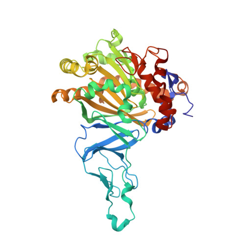
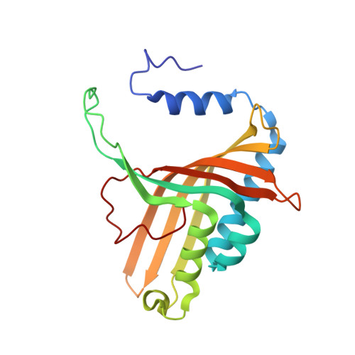
No Binding to Naphthalene Dioxygenase.

Karlsson, A.Parales, J.V.Parales, R.E.Gibson, D.T.Eklund, H.Ramaswamy, S.

(2005) J Biol Inorg Chem 10: 483

  • DOI: https://doi.org/10.1007/s00775-005-0657-1
  • Primary Citation Related Structures: 
    1UUV, 1UUW

  • PubMed Abstract: 

    Nitric oxide (NO) is commonly used as an analogue for dioxygen in structural and spectroscopic studies of oxygen binding and oxygen activation. In this study, crystallographic structures of naphthalene dioxygenase (NDO) in complex with nitric oxide are reported. In the presence of the aromatic substrate indole, NO is bound end-on to the active-site mononuclear iron of NDO. The structural observations correlate well with spectroscopic measurements of NO binding to NDO in solution. However, the end-on binding of NO is in contrast to the recently reported structure of oxygen to the active-site iron of NDO that binds side-on. While NO is a good oxygen analogue with many similarities to O(2), the different binding mode of NO to the active-site iron atom leads to different mechanistic implications. Hence, caution needs to be used in extrapolating NO as an analogue to O(2) binding.


  • Organizational Affiliation
    • Department of Molecular Biology, Biomedical Center, Swedish University of Agricultural Sciences, 75124 Uppsala, Sweden.

Macromolecule Content 

  • Total Structure Weight: 73.09 kDa 
  • Atom Count: 5,453 
  • Modeled Residue Count: 640 
  • Deposited Residue Count: 643 
  • Unique protein chains: 2

Macromolecules

Find similar proteins by:|  3D Structure
Entity ID: 1
MoleculeChains  Sequence LengthOrganismDetailsImage
NAPHTHALENE 1,2-DIOXYGENASE ALPHA SUBUNIT449Pseudomonas putidaMutation(s): 0 
EC: 1.14.12.12
UniProt
Find proteins for P0A110 (Pseudomonas putida)
Explore P0A110 
Go to UniProtKB:  P0A110
Entity Groups
Sequence Clusters30% Identity50% Identity70% Identity90% Identity95% Identity100% Identity
UniProt GroupP0A110
Sequence Annotations
Expand
Reference Sequence
Find similar proteins by:|  3D Structure
Entity ID: 2
MoleculeChains  Sequence LengthOrganismDetailsImage
NAPHTHALENE 1,2-DIOXYGENASE BETA SUBUNIT194Pseudomonas putidaMutation(s): 0 
EC: 1.14.12.12
UniProt
Find proteins for P0A112 (Pseudomonas putida)
Explore P0A112 
Go to UniProtKB:  P0A112
Entity Groups
Sequence Clusters30% Identity50% Identity70% Identity90% Identity95% Identity100% Identity
UniProt GroupP0A112
Sequence Annotations
Expand
Reference Sequence

Experimental Data & Validation

Experimental Data

  • Method: X-RAY DIFFRACTION
  • Resolution: 2.30 Å
  • R-Value Free:  0.194 (Depositor), 0.191 (DCC) 
  • R-Value Work:  0.198 (Depositor), 0.191 (DCC) 
Space Group: H 3 2
Unit Cell:
Length ( Å )Angle ( ˚ )
a = 140.556α = 90
b = 140.556β = 90
c = 210.288γ = 120
Software Package:
Software NamePurpose
REFMACrefinement
MOSFLMdata reduction
SCALAdata scaling
AMoREphasing

Structure Validation

View Full Validation Report


Entry History 

Deposition Data

Revision History  (Full details and data files)

  • Version 1.0: 2005-02-09
    Type: Initial release
  • Version 1.1: 2011-05-08
    Changes: Version format compliance
  • Version 1.2: 2011-07-13
    Changes: Version format compliance
  • Version 1.3: 2023-12-13
    Changes: Data collection, Database references, Derived calculations, Other, Refinement description