1UUJ | pdb_00001uuj

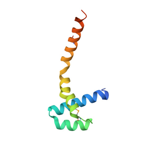
N-terminal domain of Lissencephaly-1 protein (Lis-1)


Experimental Data Snapshot

  • Method: X-RAY DIFFRACTION
  • Resolution: 1.75 Å
  • R-Value Free: 
    0.246 (Depositor), 0.254 (DCC) 
  • R-Value Work: 
    0.190 (Depositor), 0.203 (DCC) 
  • R-Value Observed: 
    0.192 (Depositor) 

wwPDB Validation 3D Report Full Report

Validation slider image for 1UUJ

This is version 1.4 of the entry. See complete history. 

Literature

The Structure of the N-Terminal Domain of the Product of the Lissencephaly Gene Lis1 and its Functional Implications

Kim, M.H., Cooper, D.R., Oleksy, A., Devedjiev, Y., Derewenda, U., Reiner, O., Otlewski, J., Derewenda, Z.S.

(2004) Structure 12: 987

  • DOI: https://doi.org/10.1016/j.str.2004.03.024
  • Primary Citation Related Structures: 
    1UUJ

  • PubMed Abstract: 

    Mutations in the Lis1 gene result in lissencephaly (smooth brain), a debilitating developmental syndrome caused by the impaired ability of postmitotic neurons to migrate to their correct destination in the cerebral cortex. Sequence similarities suggest that the LIS1 protein contains a C-terminal seven-blade beta-propeller domain, while the structure of the N-terminal fragment includes the LisH (Lis-homology) motif, a pattern found in over 100 eukaryotic proteins with a hitherto unknown function. We present the 1.75 A resolution crystal structure of the N-terminal domain of mouse LIS1, and we show that the LisH motif is a novel, thermodynamically very stable dimerization domain. The structure explains the molecular basis of a low severity form of lissencephaly.


  • Organizational Affiliation: 
    • Department of Molecular Physiology and Biological Physics and Cancer Center, University of Virginia, Charlottesville, VA 22908, USA.

Macromolecule Content 

  • Total Structure Weight: 41.65 kDa 
  • Atom Count: 2,772 
  • Modeled Residue Count: 307 
  • Deposited Residue Count: 352 
  • Unique protein chains: 1

Macromolecules

Find similar proteins by:|  3D Structure
Entity ID: 1
MoleculeChains  Sequence LengthOrganismDetailsImage
PLATELET-ACTIVATING FACTOR ACETYLHYDROLASE IB ALPHA SUBUNIT
A, B, C, D
88Mus musculusMutation(s): 0 
UniProt
Find proteins for P63005 (Mus musculus)
Explore P63005 
Go to UniProtKB:  P63005
Entity Groups
Sequence Clusters30% Identity50% Identity70% Identity90% Identity95% Identity100% Identity
UniProt GroupP63005
Sequence Annotations
Expand
Reference Sequence

Small Molecules

Modified Residues  1 Unique
IDChains TypeFormula2D DiagramParent
MSE
Query on MSE
A, B, C, D
L-PEPTIDE LINKINGC5 H11 N O2 SeMET

Experimental Data & Validation

Experimental Data

  • Method: X-RAY DIFFRACTION
  • Resolution: 1.75 Å
  • R-Value Free:  0.246 (Depositor), 0.254 (DCC) 
  • R-Value Work:  0.190 (Depositor), 0.203 (DCC) 
  • R-Value Observed: 0.192 (Depositor) 
Space Group: P 21 21 2
Unit Cell:
Length ( Å )Angle ( ˚ )
a = 62.988α = 90
b = 111.753β = 90
c = 47.397γ = 90
Software Package:
Software NamePurpose
REFMACrefinement
HKL-2000data reduction
HKL-2000data scaling
SOLVEphasing
SHARPphasing
ARP/wARPphasing

Structure Validation

View Full Validation Report


Entry History 

Deposition Data

Revision History  (Full details and data files)

  • Version 1.0: 2004-07-29
    Type: Initial release
  • Version 1.1: 2011-07-13
    Changes: Advisory, Refinement description, Version format compliance
  • Version 1.2: 2017-06-28
    Changes: Refinement description
  • Version 1.3: 2019-05-29
    Changes: Data collection, Derived calculations, Experimental preparation
  • Version 1.4: 2024-10-23
    Changes: Data collection, Database references, Other, Structure summary