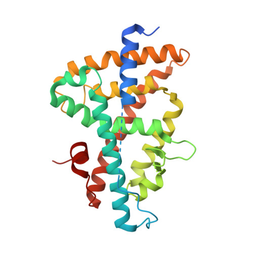
1UPV

Crystal structure of the human Liver X receptor beta ligand binding domain in complex with a synthetic agonist


Experimental Data Snapshot

  • Method: X-RAY DIFFRACTION
  • Resolution: 2.10 Å
  • R-Value Free: 0.274 
  • R-Value Work: 0.215 

Starting Model: experimental
View more details

wwPDB Validation   3D Report Full Report


This is version 1.4 of the entry. See complete history


Literature

Crystal Structure of the Human Liver X Receptor Beta Ligand-Binding Domain in Complex with a Synthetic Agonist

Hoerer, S.Schmid, A.Heckel, A.Budzinski, R.M.Nar, H.

(2003) J Mol Biol 334: 853

  • DOI: https://doi.org/10.1016/j.jmb.2003.10.033
  • Primary Citation of Related Structures:  
    1UPV, 1UPW

  • PubMed Abstract: 

    LXRbeta belongs to the nuclear hormone receptor superfamily of ligand-activated transcription factors. Its natural ligands are supposed to be oxidised derivatives of cholesterol. Stimulation of LXRbeta by agonists activates a number of genes that are involved in the regulation of lipid metabolism and cholesterol efflux from cells. Therefore, LXRbeta may represent a novel therapeutic target for the treatment of dyslipidemia and atherosclerosis.Here, we report the X-ray crystal structure of the LXRbeta ligand-binding domain in complex with a synthetic agonist, T-0901317. This compound occupies the ligand-binding pocket of the receptor, forms numerous lipophilic contacts with the protein and one crucial hydrogen bond to His435 and stabilises the agonist conformation of the receptor ligand-binding domain. The recruitment of the AF2-region of the protein is not achieved via direct polar interactions of the ligand with protein side-chains of this helical segment, but rather via few hydrophobic contacts and probably more importantly via indirect effects involving the pre-orientation of side-chains that surround the ligand-binding pocket and form the interface to the AF2-helix. On the basis of these results we propose a binding mode and a mechanism of action for the putative natural ligands, oxidised derivatives of cholesterol.


  • Organizational Affiliation

    Department of Lead Discovery, Boehringer Ingelheim Pharma GmbH & Co. KG, Birkendorfer Strasse 65, D-88397 Biberach/Riss, Germany.


Macromolecules
Find similar proteins by:  (by identity cutoff)  |  3D Structure
Entity ID: 1
MoleculeChains Sequence LengthOrganismDetailsImage
OXYSTEROLS RECEPTOR LXR-BETA257Homo sapiensMutation(s): 0 
UniProt & NIH Common Fund Data Resources
Find proteins for P55055 (Homo sapiens)
Go to UniProtKB:  P55055
PHAROS:  P55055
GTEx:  ENSG00000131408 
Sequence Annotations
Expand
  • Reference Sequence
Small Molecules
Ligands 1 Unique
IDChains Name / Formula / InChI Key2D Diagram3D Interactions
444
Query on 444

Download Ideal Coordinates CCD File 
B [auth A]N-(2,2,2-TRIFLUOROETHYL)-N-{4-[2,2,2-TRIFLUORO-1-HYDROXY-1-(TRIFLUOROMETHYL)ETHYL]PHENYL}BENZENESULFONAMIDE
C17 H12 F9 N O3 S
SGIWFELWJPNFDH-UHFFFAOYSA-N
Binding Affinity Annotations 
IDSourceBinding Affinity
444 BindingDB:  1UPV Ki: min: 17, max: 41 (nM) from 2 assay(s)
Kd: 103 (nM) from 1 assay(s)
IC50: min: 9, max: 2.60e+4 (nM) from 9 assay(s)
EC50: min: 0.11, max: 800 (nM) from 35 assay(s)
Experimental Data & Validation

Experimental Data

  • Method: X-RAY DIFFRACTION
  • Resolution: 2.10 Å
  • R-Value Free: 0.274 
  • R-Value Work: 0.215 
  • Space Group: C 1 2 1
Unit Cell:
Length ( Å )Angle ( ˚ )
a = 141.8α = 90
b = 43.2β = 105.8
c = 51.8γ = 90
Software Package:
Software NamePurpose
REFMACrefinement
MOSFLMdata reduction
SCALAdata scaling
AMoREphasing

Structure Validation

View Full Validation Report


Entry History 

Deposition Data

Revision History  (Full details and data files)

  • Version 1.0: 2004-10-20
    Type: Initial release
  • Version 1.1: 2011-05-08
    Changes: Version format compliance
  • Version 1.2: 2011-07-13
    Changes: Version format compliance
  • Version 1.3: 2018-03-28
    Changes: Source and taxonomy
  • Version 1.4: 2023-12-13
    Changes: Data collection, Database references, Derived calculations, Refinement description