1UP6 | pdb_00001up6

Structure of the 6-phospho-beta glucosidase from Thermotoga maritima at 2.55 Angstrom resolution in the tetragonal form with manganese, NAD+ and glucose-6-phosphate


Experimental Data Snapshot

  • Method: X-RAY DIFFRACTION
  • Resolution: 2.55 Å
  • R-Value Free: 
    0.250 (Depositor), 0.296 (DCC) 
  • R-Value Work: 
    0.200 (Depositor), 0.264 (DCC) 
  • R-Value Observed: 
    0.202 (Depositor) 

Starting Model: experimental
View more details

wwPDB Validation 3D Report Full Report

Validation slider image for 1UP6

Ligand Structure Quality Assessment 


This is version 1.5 of the entry. See complete history

Literature

An Unusual Mechanism of Glycoside Hydrolysis Involving Redox and Elimination Steps by a Family 4 Beta-Glycosidase from Thermotoga Maritima.

Yip, V.L.Varrot, A.Davies, G.J.Rajan, S.S.Yang, X.Thompson, J.Anderson, W.F.Withers, S.G.

(2004) J Am Chem Soc 126: 8354

  • DOI: https://doi.org/10.1021/ja047632w
  • Primary Citation Related Structures: 
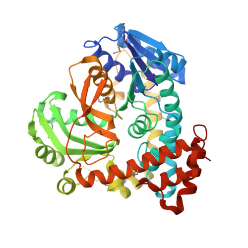
    1UP6

  • PubMed Abstract: 

    Among the numerous well-characterized families of glycosidases, family 4 appears to be the anomaly, requiring both catalytic NAD+ and a divalent metal for activity. The unusual cofactor requirement prompted the proposal of a mechanism involving key NAD+-mediated redox steps as well as elimination of the glycosidic oxygen. Primary kinetic isotope effects for the 2- and 3-deutero substrate analogues, isotopic exchange with solvent, and structural analysis of a 6-phospho-beta-glucosidase, BglT (E.C. 3.2.1.6), provided evidence in support of the proposed mechanism, which has striking resemblances to that of the sugar dehydratases. Furthermore, analysis of the stereochemical outcome indicated that family 4 enzymes are retaining glycosidases.


  • Organizational Affiliation
    • Department of Chemistry, University of British Columbia, Vancouver, BC, Canada V6T 1Z1.

Macromolecule Content 

  • Total Structure Weight: 388.45 kDa 
  • Atom Count: 26,917 
  • Modeled Residue Count: 3,222 
  • Deposited Residue Count: 3,328 
  • Unique protein chains: 1

Macromolecules

Find similar proteins by:|  3D Structure
Entity ID: 1
MoleculeChains  Sequence LengthOrganismDetailsImage
6-PHOSPHO-BETA-GLUCOSIDASE
A, B, C, D, E
A, B, C, D, E, F, G, H
416Thermotoga maritima MSB8Mutation(s): 0 
EC: 3.2.1.6 (PDB Primary Data), 3.2.1.86 (UniProt)
UniProt
Find proteins for Q9X108 (Thermotoga maritima (strain ATCC 43589 / DSM 3109 / JCM 10099 / NBRC 100826 / MSB8))
Explore Q9X108 
Go to UniProtKB:  Q9X108
Entity Groups
Sequence Clusters30% Identity50% Identity70% Identity90% Identity95% Identity100% Identity
UniProt GroupQ9X108
Sequence Annotations
Expand
Reference Sequence

Small Molecules

Ligands 4 Unique
IDChains Name / Formula / InChI Key2D Diagram3D Interactions
NAD

Query on NAD


Download:Ideal Coordinates CCD File
I [auth A],
O [auth C],
T [auth G]
NICOTINAMIDE-ADENINE-DINUCLEOTIDE
C21 H27 N7 O14 P2
BAWFJGJZGIEFAR-NNYOXOHSSA-N
G6P

Query on G6P


Download:Ideal Coordinates CCD File
K [auth A],
Q [auth C]
6-O-phosphono-alpha-D-glucopyranose
C6 H13 O9 P
NBSCHQHZLSJFNQ-DVKNGEFBSA-N
SO4

Query on SO4


Download:Ideal Coordinates CCD File
N [auth B],
R [auth D],
S [auth E],
V [auth G]
SULFATE ION
O4 S
QAOWNCQODCNURD-UHFFFAOYSA-L
MN

Query on MN


Download:Ideal Coordinates CCD File
J [auth A],
L [auth A],
M [auth A],
P [auth C],
U [auth G]
MANGANESE (II) ION
Mn
WAEMQWOKJMHJLA-UHFFFAOYSA-N
Modified Residues  1 Unique
IDChains TypeFormula2D DiagramParent
MSE
Query on MSE
A, B, C, D, E
A, B, C, D, E, F, G, H
L-PEPTIDE LINKINGC5 H11 N O2 SeMET

Experimental Data & Validation

Experimental Data

  • Method: X-RAY DIFFRACTION
  • Resolution: 2.55 Å
  • R-Value Free:  0.250 (Depositor), 0.296 (DCC) 
  • R-Value Work:  0.200 (Depositor), 0.264 (DCC) 
  • R-Value Observed: 0.202 (Depositor) 
Space Group: P 43 21 2
Unit Cell:
Length ( Å )Angle ( ˚ )
a = 179.438α = 90
b = 179.438β = 90
c = 282.911γ = 90
Software Package:
Software NamePurpose
REFMACrefinement
DENZOdata reduction
SCALEPACKdata scaling
AMoREphasing

Structure Validation

View Full Validation Report


Ligand Structure Quality Assessment 


Entry History 

Deposition Data

Revision History  (Full details and data files)

  • Version 1.0: 2004-08-02
    Type: Initial release
  • Version 1.1: 2011-05-08
    Changes: Version format compliance
  • Version 1.2: 2011-07-13
    Changes: Version format compliance
  • Version 1.3: 2020-07-29
    Type: Remediation
    Reason: Carbohydrate remediation
    Changes: Advisory, Data collection, Derived calculations, Other, Structure summary
  • Version 1.4: 2023-12-13
    Changes: Advisory, Data collection, Database references, Refinement description, Structure summary
  • Version 1.5: 2024-10-09
    Changes: Structure summary