1UOZ | pdb_00001uoz

Structure of the endoglucanase Cel6 from Mycobacterium tuberculosis in complex with thiocellopentaose at 1.1 angstrom


Experimental Data Snapshot

  • Method: X-RAY DIFFRACTION
  • Resolution: 1.10 Å
  • R-Value Free: 
    0.130 (Depositor), 0.162 (DCC) 
  • R-Value Work: 
    0.112 (Depositor) 
  • R-Value Observed: 
    0.113 (Depositor) 

Starting Model: experimental
View more details

wwPDB Validation 3D Report Full Report

Validation slider image for 1UOZ

This is version 2.2 of the entry. See complete history

Literature

Mycobacterium Tuberculosis Strains Possess Functional Cellulases.

Varrot, A.Leydier, S.Pell, G.Macdonald, J.M.Stick, R.V.Henrissat, B.Gilbert, H.J.Davies, G.J.

(2005) J Biological Chem 280: 20181

  • DOI: https://doi.org/10.1074/jbc.C500142200
  • Primary Citation Related Structures: 
    1UOZ, 1UP0, 1UP2, 1UP3

  • PubMed Abstract: 

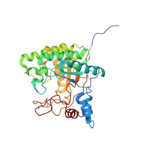
    The genomes of various Mycobacterium tuberculosis strains encode proteins that do not appear to play a role in the growth or survival of the bacterium in its mammalian host, including some implicated in plant cell wall breakdown. Here we show that M. tuberculosis H37Rv does indeed possess a functional cellulase. The x-ray crystal structure of this enzyme, in ligand complex forms, from 1.9 to 1.1A resolution, reveals a highly conserved substrate-binding cleft, which affords similar, and unusual, distortion of the substrate at the catalytic center. The endoglucanase activity, together with the existence of a putative membrane-associated crystalline polysaccharide-binding protein, may reflect the ancestral soil origin of the Mycobacterium or hint at a previously unconsidered environmental niche.


  • Organizational Affiliation
    • York Structural Biology Laboratory, Department of Chemistry, University of York, York, Y010 5YW, United Kingdom.

Macromolecule Content 

  • Total Structure Weight: 34.13 kDa 
  • Atom Count: 2,855 
  • Modeled Residue Count: 300 
  • Deposited Residue Count: 315 
  • Unique protein chains: 1

Macromolecules

Find similar proteins by:|  3D Structure
Entity ID: 1
MoleculeChains  Sequence LengthOrganismDetailsImage
PUTATIVE CELLULASE315Mycobacterium tuberculosis H37RvMutation(s): 0 
EC: 3.2.1
UniProt
Find proteins for Q79G13 (Mycobacterium tuberculosis (strain ATCC 25618 / H37Rv))
Explore Q79G13 
Go to UniProtKB:  Q79G13
Entity Groups
Sequence Clusters30% Identity50% Identity70% Identity90% Identity95% Identity100% Identity
UniProt GroupQ79G13
Sequence Annotations
Expand
Reference Sequence

Oligosaccharides

Help  
Entity ID: 2
MoleculeChains Length2D Diagram GlycosylationD Interactions
alpha-D-glucopyranose-(1-4)-1,4-dithio-beta-D-glucopyranose-(1-4)-1,4-dithio-beta-D-glucopyranose
B
3N/AN/A
Glycosylation Resources
GlyTouCan: G10396JO
GlyCosmos: G10396JO

Experimental Data & Validation

Experimental Data

  • Method: X-RAY DIFFRACTION
  • Resolution: 1.10 Å
  • R-Value Free:  0.130 (Depositor), 0.162 (DCC) 
  • R-Value Work:  0.112 (Depositor) 
  • R-Value Observed: 0.113 (Depositor) 
Space Group: P 21 21 2
Unit Cell:
Length ( Å )Angle ( ˚ )
a = 61.94α = 90
b = 92.627β = 90
c = 46.677γ = 90
Software Package:
Software NamePurpose
REFMACrefinement
MOSFLMdata reduction
SCALAdata scaling
AMoREphasing

Structure Validation

View Full Validation Report


Entry History 

Deposition Data

Revision History  (Full details and data files)

  • Version 1.0: 2004-11-18
    Type: Initial release
  • Version 1.1: 2011-05-08
    Changes: Version format compliance
  • Version 1.2: 2011-07-13
    Changes: Version format compliance
  • Version 2.0: 2020-07-29
    Type: Remediation
    Reason: Carbohydrate remediation
    Changes: Advisory, Atomic model, Data collection, Derived calculations, Other, Structure summary
  • Version 2.1: 2023-12-13
    Changes: Data collection, Database references, Refinement description, Structure summary
  • Version 2.2: 2024-10-16
    Changes: Structure summary