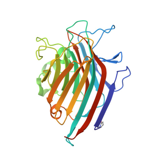
1UKG

Pterocarps angolensis lectin PAL in complex with methyl-alpha-mannose


Experimental Data Snapshot

  • Method: X-RAY DIFFRACTION
  • Resolution: 1.70 Å
  • R-Value Free: 0.215 
  • R-Value Work: 0.189 
  • R-Value Observed: 0.191 

wwPDB Validation   3D Report Full Report


Ligand Structure Quality Assessment 


This is version 2.3 of the entry. See complete history


Literature

Structural Basis of Oligomannose Recognition by the Pterocarpus angolensis Seed Lectin

Loris, R.Van Walle, I.De Greve, H.Beeckmans, S.Deboeck, F.Wyns, L.Bouckaert, J.

(2004) J Mol Biol 335: 1227-1240

  • DOI: https://doi.org/10.1016/j.jmb.2003.11.043
  • Primary Citation of Related Structures:  
    1Q8O, 1Q8P, 1Q8Q, 1Q8S, 1Q8V, 1UKG

  • PubMed Abstract: 

    The crystal structure of a Man/Glc-specific lectin from the seeds of the bloodwood tree (Pterocarpus angolensis), a leguminous plant from central Africa, has been determined in complex with mannose and five manno-oligosaccharides. The lectin contains a classical mannose-specificity loop, but its metal-binding loop resembles that of lectins of unrelated specificity from Ulex europaeus and Maackia amurensis. As a consequence, the interactions with mannose in the primary binding site are conserved, but details of carbohydrate-binding outside the primary binding site differ from those seen in the equivalent carbohydrate complexes of concanavalin A. These observations explain the differences in their respective fine specificity profiles for oligomannoses. While Man(alpha1-3)Man and Man(alpha1-3)[Man(alpha1-6)]Man bind to PAL in low-energy conformations identical with that of ConA, Man(alpha1-6)Man is required to adopt a different conformation. Man(alpha1-2)Man can bind only in a single binding mode, in sharp contrast to ConA, which creates a higher affinity for this disaccharide by allowing two binding modes.


  • Organizational Affiliation

    Laboratorium voor Ultrastructuur, Instituut voor Moleculaire Biologie, Building E, Vrije Universiteit Brussel and Vlaams Instituut voor Biotechnologie, Pleinlaan 2, B-1050 Brussel, Belgium. reloris@vub.ac.be


Macromolecules
Find similar proteins by:  (by identity cutoff)  |  3D Structure
Entity ID: 1
MoleculeChains Sequence LengthOrganismDetailsImage
lectin
A, B
252Pterocarpus angolensisMutation(s): 0 
UniProt
Find proteins for Q8GSD2 (Pterocarpus angolensis)
Explore Q8GSD2 
Go to UniProtKB:  Q8GSD2
Entity Groups  
Sequence Clusters30% Identity50% Identity70% Identity90% Identity95% Identity100% Identity
UniProt GroupQ8GSD2
Sequence Annotations
Expand
  • Reference Sequence
Small Molecules
Modified Residues  1 Unique
IDChains TypeFormula2D DiagramParent
PCA
Query on PCA
A, B
L-PEPTIDE LINKINGC5 H7 N O3GLN
Experimental Data & Validation

Experimental Data

  • Method: X-RAY DIFFRACTION
  • Resolution: 1.70 Å
  • R-Value Free: 0.215 
  • R-Value Work: 0.189 
  • R-Value Observed: 0.191 
  • Space Group: P 21 21 21
Unit Cell:
Length ( Å )Angle ( ˚ )
a = 56.82α = 90
b = 83.67β = 90
c = 123γ = 90
Software Package:
Software NamePurpose
DENZOdata reduction
SCALEPACKdata scaling
AMoREphasing
CNSrefinement

Structure Validation

View Full Validation Report


Ligand Structure Quality Assessment 


Entry History 

Deposition Data

Revision History  (Full details and data files)

  • Version 1.0: 2004-02-10
    Type: Initial release
  • Version 1.1: 2008-04-27
    Changes: Version format compliance
  • Version 1.2: 2011-07-13
    Changes: Version format compliance
  • Version 2.0: 2019-12-25
    Changes: Data collection, Database references, Derived calculations, Polymer sequence
  • Version 2.1: 2020-07-29
    Type: Remediation
    Reason: Carbohydrate remediation
    Changes: Data collection, Derived calculations, Structure summary
  • Version 2.2: 2023-12-27
    Changes: Data collection, Database references, Structure summary
  • Version 2.3: 2024-11-13
    Changes: Structure summary