1UJW

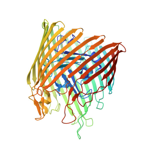
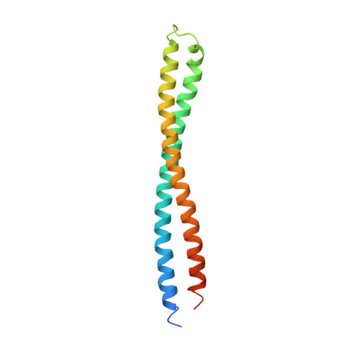
Structure of the complex between BtuB and Colicin E3 Receptor binding domain


Experimental Data Snapshot

  • Method: X-RAY DIFFRACTION
  • Resolution: 2.75 Å
  • R-Value Free: 0.293 
  • R-Value Work: 0.244 
  • R-Value Observed: 0.247 

Starting Model: experimental
View more details

wwPDB Validation   3D Report Full Report


Ligand Structure Quality Assessment 


This is version 2.1 of the entry. See complete history


Literature

The structure of BtuB with bound colicin E3 R-domain implies a translocon

Kurisu, G.Zakharov, S.D.Zhalnina, M.V.Bano, S.Eroukova, V.Y.Rokitskaya, T.I.Antonenko, Y.N.Wiener, M.C.Cramer, W.A.

(2003) Nat Struct Biol 10: 948-954

  • DOI: https://doi.org/10.1038/nsb997
  • Primary Citation of Related Structures:  
    1UJW

  • PubMed Abstract: 

    Cellular import of colicin E3 is initiated by the Escherichia coli outer membrane cobalamin transporter, BtuB. The 135-residue 100-A coiled-coil receptor-binding domain (R135) of colicin E3 forms a 1:1 complex with BtuB whose structure at a resolution of 2.75 A is reported. Binding of R135 to the BtuB extracellular surface (DeltaG(o) = -12 kcal mol(-1)) is mediated by 27 residues of R135 near the coiled-coil apex. Formation of the R135-BtuB complex results in unfolding of R135 N- and C-terminal ends, inferred to be important for unfolding of the colicin T-domain. Small conformational changes occur in the BtuB cork and barrel domains but are insufficient to form a translocation channel. The absence of a channel and the peripheral binding of R135 imply that BtuB serves to bind the colicin, and that the coiled-coil delivers the colicin to a neighboring outer membrane protein for translocation, thus forming a colicin translocon. The translocator was concluded to be OmpF from the occlusion of OmpF channels by colicin E3.


  • Organizational Affiliation

    Department of Biological Sciences, Purdue University, Lilly Hall of Life Sciences, 915 W. State St., West Lafayette, Indiana 47907-1392, USA.


Macromolecules
Find similar proteins by:  (by identity cutoff)  |  3D Structure
Entity ID: 1
MoleculeChains Sequence LengthOrganismDetailsImage
Vitamin B12 receptor594Escherichia coliMutation(s): 0 
Membrane Entity: Yes 
UniProt
Find proteins for P06129 (Escherichia coli (strain K12))
Explore P06129 
Go to UniProtKB:  P06129
Entity Groups  
Sequence Clusters30% Identity50% Identity70% Identity90% Identity95% Identity100% Identity
UniProt GroupP06129
Sequence Annotations
Expand
  • Reference Sequence
Find similar proteins by:  (by identity cutoff)  |  3D Structure
Entity ID: 2
MoleculeChains Sequence LengthOrganismDetailsImage
Colicin E3135Escherichia coliMutation(s): 0 
UniProt
Find proteins for P00646 (Escherichia coli)
Explore P00646 
Go to UniProtKB:  P00646
Entity Groups  
Sequence Clusters30% Identity50% Identity70% Identity90% Identity95% Identity100% Identity
UniProt GroupP00646
Sequence Annotations
Expand
  • Reference Sequence
Small Molecules
Ligands 5 Unique
IDChains Name / Formula / InChI Key2D Diagram3D Interactions
GP1
Query on GP1

Download Ideal Coordinates CCD File 
C [auth A],
D [auth A]
2-amino-2-deoxy-1-O-phosphono-alpha-D-glucopyranose
C6 H14 N O8 P
YMJBYRVFGYXULK-QZABAPFNSA-N
LIM
Query on LIM

Download Ideal Coordinates CCD File 
E [auth A],
N [auth A]
3-OXO-PENTADECANOIC ACID
C15 H28 O3
CJTNJZWHYGHVCD-UHFFFAOYSA-N
LDA
Query on LDA

Download Ideal Coordinates CCD File 
F [auth A]
G [auth A]
H [auth A]
I [auth A]
J [auth A]
F [auth A],
G [auth A],
H [auth A],
I [auth A],
J [auth A],
K [auth A],
L [auth A]
LAURYL DIMETHYLAMINE-N-OXIDE
C14 H31 N O
SYELZBGXAIXKHU-UHFFFAOYSA-N
AAE
Query on AAE

Download Ideal Coordinates CCD File 
M [auth A]ACETOACETIC ACID
C4 H6 O3
WDJHALXBUFZDSR-UHFFFAOYSA-N
GOL
Query on GOL

Download Ideal Coordinates CCD File 
O [auth A]
P [auth A]
Q [auth A]
R [auth A]
S [auth A]
O [auth A],
P [auth A],
Q [auth A],
R [auth A],
S [auth A],
T [auth A],
U [auth A],
V [auth A]
GLYCEROL
C3 H8 O3
PEDCQBHIVMGVHV-UHFFFAOYSA-N
Experimental Data & Validation

Experimental Data

  • Method: X-RAY DIFFRACTION
  • Resolution: 2.75 Å
  • R-Value Free: 0.293 
  • R-Value Work: 0.244 
  • R-Value Observed: 0.247 
  • Space Group: P 21 21 21
Unit Cell:
Length ( Å )Angle ( ˚ )
a = 76.933α = 90
b = 80.098β = 90
c = 233.595γ = 90
Software Package:
Software NamePurpose
REFMACrefinement
HKL-2000data reduction
HKL-2000data scaling
AMoREphasing

Structure Validation

View Full Validation Report


Ligand Structure Quality Assessment 


Entry History 

Deposition Data

Revision History  (Full details and data files)

  • Version 1.0: 2003-11-25
    Type: Initial release
  • Version 1.1: 2008-04-27
    Changes: Version format compliance
  • Version 1.2: 2011-07-13
    Changes: Advisory, Version format compliance
  • Version 2.0: 2020-07-29
    Type: Remediation
    Reason: Carbohydrate remediation
    Changes: Atomic model, Data collection, Derived calculations, Structure summary
  • Version 2.1: 2023-10-25
    Changes: Data collection, Database references, Derived calculations, Refinement description, Structure summary