1UHH | pdb_00001uhh

Crystal structure of cp-aequorin


Experimental Data Snapshot

  • Method: X-RAY DIFFRACTION
  • Resolution: 1.80 Å
  • R-Value Free: 
    0.190 (Depositor), 0.204 (DCC) 
  • R-Value Work: 
    0.142 (Depositor), 0.155 (DCC) 
  • R-Value Observed: 
    0.145 (Depositor) 

Starting Model: experimental
View more details

wwPDB Validation 3D Report Full Report

Validation slider image for 1UHH

Ligand Structure Quality Assessment 


This is version 1.5 of the entry. See complete history

Literature

The crystal structures of semi-synthetic aequorins

Toma, S.Chong, K.T.Nakagawa, A.Teranishi, K.Inouye, S.Shimomura, O.

(2005) Protein Sci 14: 409-416

  • DOI: https://doi.org/10.1110/ps.041067805
  • Primary Citation Related Structures: 
    1UHH, 1UHI, 1UHJ, 1UHK

  • PubMed Abstract: 

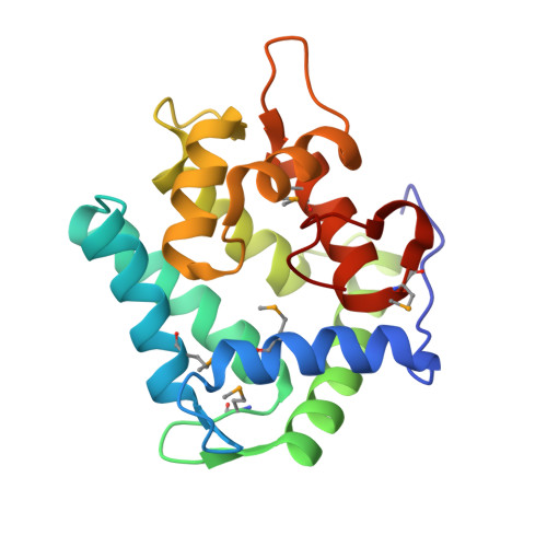
    The photoprotein aequorin emits light by an intramolecular reaction in the presence of a trace amount of Ca(2+). Semi-synthetic aequorins, produced by replacing the coelenterazine moiety in aequorin with the analogues of coelenterazine, show widely different sensitivities to Ca(2+). To understand the structural basis of the Ca(2+)-sensitivity, we determined the crystal structures of four semi-synthetic aequorins (cp-, i-, br- and n-aequorins) at resolutions of 1.6-1.8 A. In general, the protein structures of these semi-synthetic aequorins are almost identical to native aequorin. Of the four EF-hand domains in the molecule, EF-hand II does not bind Ca(2+), and the loop of EF-hand IV is clearly deformed. It is most likely that the binding of Ca(2+) with EF-hands I and III triggers luminescence. Although little difference was found in the overall structures of aequorins investigated, some significant differences were found in the interactions between the substituents of coelenterazine moiety and the amino acid residues in the binding pocket. The coelenterazine moieties in i-, br-, and n-aequorins have bulky 2-substitutions, which can interfere with the conformational changes of protein structure that follow the binding of Ca(2+) to aequorin. In cp-aequorin, the cyclopentylmethyl group that substitutes for the original 8-benzyl group does not interact hydrophobically with the protein part, giving the coelenterazine moiety more conformational freedom to promote the light-emitting reaction. The differences of various semi-synthetic aequorins in Ca(2+)-sensitivity and reaction rate are explained by the capability of the involved groups and structures to undergo conformational changes in response to the Ca(2+)-binding.


  • Organizational Affiliation
    • Institute for Protein Research, Osaka University, 3-2 Yamadaoka, Suita, Osaka 565-0871, Japan.

Macromolecule Content 

  • Total Structure Weight: 44.68 kDa 
  • Atom Count: 3,652 
  • Modeled Residue Count: 378 
  • Deposited Residue Count: 382 
  • Unique protein chains: 1

Macromolecules

Find similar proteins by:|  3D Structure
Entity ID: 1
MoleculeChains  Sequence LengthOrganismDetailsImage
Aequorin 2
A, B
191Aequorea victoriaMutation(s): 5 
UniProt
Find proteins for P02592 (Aequorea victoria)
Explore P02592 
Go to UniProtKB:  P02592
Entity Groups
Sequence Clusters30% Identity50% Identity70% Identity90% Identity95% Identity100% Identity
UniProt GroupP02592
Sequence Annotations
Expand
Reference Sequence

Small Molecules

Ligands 1 Unique
IDChains Name / Formula / InChI Key2D Diagram3D Interactions
CZP

Query on CZP


Download:Ideal Coordinates CCD File
C [auth A],
D [auth B]
(8R)-8-(CYCLOPENTYLMETHYL)-2-HYDROPEROXY-2-(4-HYDROXYBENZYL)-6-(4-HYDROXYPHENYL)-7,8-DIHYDROIMIDAZO[1,2-A]PYRAZIN-3(2H) -ONE
C25 H27 N3 O5
XOSFCMOTHZJUQK-OFVILXPXSA-N
Modified Residues  1 Unique
IDChains TypeFormula2D DiagramParent
MSE
Query on MSE
A, B
L-PEPTIDE LINKINGC5 H11 N O2 SeMET

Experimental Data & Validation

Experimental Data

  • Method: X-RAY DIFFRACTION
  • Resolution: 1.80 Å
  • R-Value Free:  0.190 (Depositor), 0.204 (DCC) 
  • R-Value Work:  0.142 (Depositor), 0.155 (DCC) 
  • R-Value Observed: 0.145 (Depositor) 
Space Group: P 1
Unit Cell:
Length ( Å )Angle ( ˚ )
a = 34.198α = 72.2
b = 57.274β = 80
c = 58.191γ = 68.4
Software Package:
Software NamePurpose
REFMACrefinement
d*TREKdata reduction
d*TREKdata scaling
MOLREPphasing

Structure Validation

View Full Validation Report


Ligand Structure Quality Assessment 


Entry History 

Deposition Data

Revision History  (Full details and data files)

  • Version 1.0: 2005-02-08
    Type: Initial release
  • Version 1.1: 2008-04-27
    Changes: Version format compliance
  • Version 1.2: 2011-07-13
    Changes: Version format compliance
  • Version 1.3: 2023-10-25
    Changes: Data collection, Database references, Derived calculations, Refinement description, Structure summary
  • Version 1.4: 2023-11-15
    Changes: Data collection
  • Version 1.5: 2024-10-30
    Changes: Structure summary