1UDI | pdb_00001udi

NUCLEOTIDE MIMICRY IN THE CRYSTAL STRUCTURE OF THE URACIL-DNA GLYCOSYLASE-URACIL GLYCOSYLASE INHIBITOR PROTEIN COMPLEX


Experimental Data Snapshot

  • Method: X-RAY DIFFRACTION
  • Resolution: 2.70 Å
  • R-Value Free: 
    0.276 (Depositor) 
  • R-Value Work: 
    0.188 (Depositor) 
  • R-Value Observed: 
    0.188 (Depositor) 

wwPDB Validation 3D Report Full Report

Validation slider image for 1UDI

This is version 1.3 of the entry. See complete history

Literature

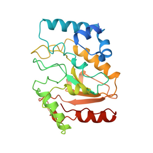
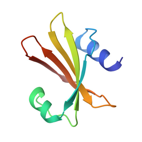
Nucleotide mimicry in the crystal structure of the uracil-DNA glycosylase-uracil glycosylase inhibitor protein complex.

Savva, R.Pearl, L.H.

(1995) Nat Struct Biol 2: 752-757

  • DOI: https://doi.org/10.1038/nsb0995-752
  • Primary Citation Related Structures: 
    1UDI

  • PubMed Abstract: 

    The Bacillus subtilis bacteriophages PBS-1 and PBS-2 protect their uracil-containing DNA by expressing an inhibitor protein (UGI) which inactivates the host uracil-DNA glycosylase (UDGase) base-excision repair enzyme. Also, PBS1/2 UGI efficiently inactivates UDGases from other biological sources, including the enzyme from herpes simplex virus type-1 (HSV-1). The crystal structure of the HSV-1 UDGase-PBS1 UGI complex at 2.7 angstrum reveals an alpha-beta-alpha sandwich structure for UGI which interacts with conserved regions of UDGase involved in DNA binding, and directly mimics protein-DNA interactions observed in the UDGase-oligonucleotide complex. The inhibitor completely blocks access to the active site of UDGase, but makes no direct contact with the uracil-binding pocket itself.


  • Organizational Affiliation
    • Department of Biochemistry and Molecular Biology, University College, London, UK.

Macromolecule Content 

  • Total Structure Weight: 36.72 kDa 
  • Atom Count: 2,519 
  • Modeled Residue Count: 310 
  • Deposited Residue Count: 327 
  • Unique protein chains: 2

Macromolecules

Find similar proteins by:|  3D Structure
Entity ID: 1
MoleculeChains  Sequence LengthOrganismDetailsImage
URACIL-DNA GLYCOSYLASEA [auth E]244Human alphaherpesvirus 1 strain 17Mutation(s): 0 
EC: 3.2.2.3 (PDB Primary Data), 3.2.2.27 (UniProt)
UniProt
Find proteins for P10186 (Human herpesvirus 1 (strain 17))
Explore P10186 
Go to UniProtKB:  P10186
Entity Groups
Sequence Clusters30% Identity50% Identity70% Identity90% Identity95% Identity100% Identity
UniProt GroupP10186
Sequence Annotations
Expand
Reference Sequence
Find similar proteins by:|  3D Structure
Entity ID: 2
MoleculeChains  Sequence LengthOrganismDetailsImage
URACIL-DNA GLYCOSYLASE INHIBITOR PROTEINB [auth I]83Takahashivirus PBS1Mutation(s): 0 
UniProt
Find proteins for P14739 (Bacillus phage PBS2)
Explore P14739 
Go to UniProtKB:  P14739
Entity Groups
Sequence Clusters30% Identity50% Identity70% Identity90% Identity95% Identity100% Identity
UniProt GroupP14739
Sequence Annotations
Expand
Reference Sequence

Experimental Data & Validation

Experimental Data

  • Method: X-RAY DIFFRACTION
  • Resolution: 2.70 Å
  • R-Value Free:  0.276 (Depositor) 
  • R-Value Work:  0.188 (Depositor) 
  • R-Value Observed: 0.188 (Depositor) 
Space Group: P 65
Unit Cell:
Length ( Å )Angle ( ˚ )
a = 144.12α = 90
b = 144.12β = 90
c = 40.95γ = 120
Software Package:
Software NamePurpose
X-PLORmodel building
X-PLORrefinement
MOSFLMdata reduction
X-PLORphasing

Structure Validation

View Full Validation Report


Entry History 

Deposition Data

Revision History  (Full details and data files)

  • Version 1.0: 1996-03-08
    Type: Initial release
  • Version 1.1: 2008-03-24
    Changes: Version format compliance
  • Version 1.2: 2011-07-13
    Changes: Version format compliance
  • Version 1.3: 2024-02-14
    Changes: Data collection, Database references, Other