1UBG | pdb_00001ubg

MsREcA-dATP complex


Experimental Data Snapshot

  • Method: X-RAY DIFFRACTION
  • Resolution: 3.50 Å
  • R-Value Free: 
    0.286 (Depositor), 0.279 (DCC) 
  • R-Value Work: 
    0.222 (Depositor), 0.218 (DCC) 
  • R-Value Observed: 
    0.222 (Depositor) 

Starting Model: experimental
View more details

wwPDB Validation 3D Report Full Report

Validation slider image for 1UBG

Ligand Structure Quality Assessment 


This is version 1.3 of the entry. See complete history

Literature

Crystal Structures of Mycobacterium smegmatis RecA and Its Nucleotide Complexes

Datta, S.Krishna, R.Ganesh, N.Chandra, N.R.Muniyappa, K.Vijayan, M.

(2003) J Bacteriol 185: 4280-4284

  • DOI: https://doi.org/10.1128/JB.185.14.4280-4284.2003
  • Primary Citation Related Structures: 
    1UBC, 1UBE, 1UBF, 1UBG

  • PubMed Abstract: 

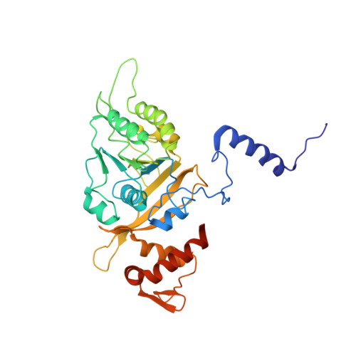
    The crystal structures of Mycobacterium smegmatis RecA (RecA(Ms)) and its complexes with ADP, ATPgammaS, and dATP show that RecA(Ms) has an expanded binding site like that in Mycobacterium tuberculosis RecA, although there are small differences between the proteins in their modes of nucleotide binding. Nucleotide binding is invariably accompanied by the movement of Gln 196, which appears to provide the trigger for transmitting the effect of nucleotide binding to the DNA-binding loops. These observations provide a framework for exploring the known properties of the RecA proteins.


  • Organizational Affiliation
    • Molecular Biophysics Unit, Indian Institute of Science, Bangalore 560 012, India.

Macromolecule Content 

  • Total Structure Weight: 37.84 kDa 
  • Atom Count: 2,486 
  • Modeled Residue Count: 330 
  • Deposited Residue Count: 349 
  • Unique protein chains: 1

Macromolecules

Find similar proteins by:|  3D Structure
Entity ID: 1
MoleculeChains  Sequence LengthOrganismDetailsImage
RecA349Mycolicibacterium smegmatisMutation(s): 0 
UniProt
Find proteins for Q59560 (Mycolicibacterium smegmatis (strain ATCC 700084 / mc(2)155))
Explore Q59560 
Go to UniProtKB:  Q59560
Entity Groups
Sequence Clusters30% Identity50% Identity70% Identity90% Identity95% Identity100% Identity
UniProt GroupQ59560
Sequence Annotations
Expand
Reference Sequence

Small Molecules

Ligands 1 Unique
IDChains Name / Formula / InChI Key2D Diagram3D Interactions
DTP

Query on DTP


Download:Ideal Coordinates CCD File
B [auth A]2'-DEOXYADENOSINE 5'-TRIPHOSPHATE
C10 H16 N5 O12 P3
SUYVUBYJARFZHO-RRKCRQDMSA-N

Experimental Data & Validation

Experimental Data

  • Method: X-RAY DIFFRACTION
  • Resolution: 3.50 Å
  • R-Value Free:  0.286 (Depositor), 0.279 (DCC) 
  • R-Value Work:  0.222 (Depositor), 0.218 (DCC) 
  • R-Value Observed: 0.222 (Depositor) 
Space Group: P 61
Unit Cell:
Length ( Å )Angle ( ˚ )
a = 102.96α = 90
b = 102.96β = 90
c = 73.578γ = 120
Software Package:
Software NamePurpose
CNSrefinement
DENZOdata reduction
SCALEPACKdata scaling
AMoREphasing

Structure Validation

View Full Validation Report


Ligand Structure Quality Assessment 


Entry History 

Deposition Data

Revision History  (Full details and data files)

  • Version 1.0: 2003-07-22
    Type: Initial release
  • Version 1.1: 2008-04-27
    Changes: Version format compliance
  • Version 1.2: 2011-07-13
    Changes: Version format compliance
  • Version 1.3: 2023-10-25
    Changes: Data collection, Database references, Derived calculations, Refinement description