1UB0 | pdb_00001ub0

Crystal Structure Analysis of Phosphomethylpyrimidine Kinase (ThiD) from Thermus Thermophilus Hb8


Experimental Data Snapshot

  • Method: X-RAY DIFFRACTION
  • Resolution: 2.05 Å
  • R-Value Free: 
    0.232 (Depositor), 0.234 (DCC) 
  • R-Value Work: 
    0.207 (Depositor), 0.209 (DCC) 

Starting Model: experimental
View more details

wwPDB Validation 3D Report Full Report

Validation slider image for 1UB0

This is version 1.4 of the entry. See complete history

Literature

Crystal structure of 4-amino-5-hydroxymethyl-2-methyl pyrimidine phosphate kinase (HMPP kinase) from Thermus thermophilus HB8.

Kambaru, A.Jos, S.Lobov, I.Bagautdinov, B.Padavattan, S.

(2025) Eur Biophys J 54: 247-256

  • DOI: https://doi.org/10.1007/s00249-025-01760-0
  • Primary Citation Related Structures: 
    1UB0

  • PubMed Abstract: 

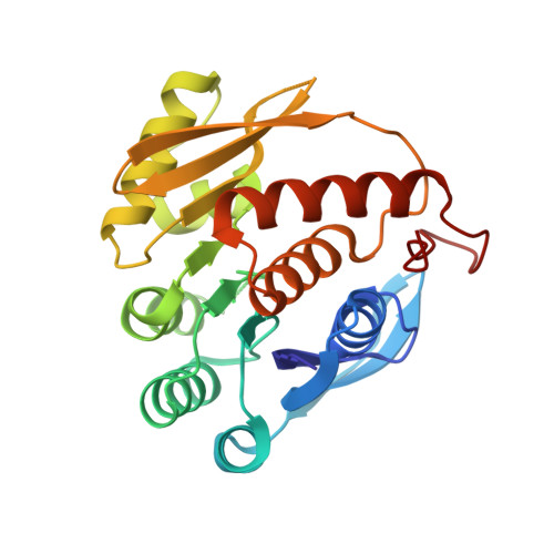
    The thiamine (vitamin B1) biosynthesis pathway is essential for most prokaryotes and some eukaryotes, including yeasts and plants. The 4-amino-5-hydroxymethyl-2-methylpyrimidine phosphate kinase (HMPP kinase), encoded by the thiD gene, catalyzes two phosphorylation reactions involving intermediates in this pathway, ultimately producing thiamine pyrophosphate, the active form of thiamine. Here, we present the crystal structure of HMPP kinase from Thermus thermophilus HB8 (TtHMPPK), resolved at a resolution of 2.05 Å. The asymmetric unit of the TtHMPPK structure includes one protomer, though it functions as a homodimer in its active form, like the HMPP kinase from Salmonella typhimurium. The TtHMPPK protomer is an α/β protein featuring nine β-sheets flanked by eight structurally conserved α-helices, which are characteristic of the ribokinase family. The structure reveals a Rossmann β-α-β motif, commonly found in nucleotide-binding proteins. Structural analysis of TtHMPPK, compared to the Salmonella typhimurium HMPP kinase, indicates that Ala16, Thr40, Gln42, Ala78, and Val105 are active site residues involved in catalysis. The structural studies suggest that TtHMPPK belongs to the ribokinase superfamily and exhibits structural similarities with an enzyme containing a Rossmann-like structural motif (RLM). This Rossmann fold enables HMPP kinase to catalyze the phosphorylation of HMPP, a critical step in thiamine production.


  • Organizational Affiliation
    • Department of Biophysics, National Institute of Mental Health and Neurosciences, Bangalore, 560029, India.

Macromolecule Content 

  • Total Structure Weight: 27.46 kDa 
  • Atom Count: 1,957 
  • Modeled Residue Count: 246 
  • Deposited Residue Count: 258 
  • Unique protein chains: 1

Macromolecules

Find similar proteins by:|  3D Structure
Entity ID: 1
MoleculeChains  Sequence LengthOrganismDetailsImage
Phosphomethylpyrimidine Kinase258Thermus thermophilusMutation(s): 0 
Gene Names: ThiD
EC: 2.7.4.7 (PDB Primary Data), 2.7.1.49 (UniProt)
UniProt
Find proteins for Q7SIA0 (Thermus thermophilus)
Explore Q7SIA0 
Go to UniProtKB:  Q7SIA0
Entity Groups
Sequence Clusters30% Identity50% Identity70% Identity90% Identity95% Identity100% Identity
UniProt GroupQ7SIA0
Sequence Annotations
Expand
Reference Sequence

Small Molecules

Ligands 3 Unique
IDChains Name / Formula / InChI Key2D Diagram3D Interactions
SO4

Query on SO4


Download:Ideal Coordinates CCD File
B [auth A]SULFATE ION
O4 S
QAOWNCQODCNURD-UHFFFAOYSA-L
GOL

Query on GOL


Download:Ideal Coordinates CCD File
E [auth A],
F [auth A],
G [auth A],
H [auth A]
GLYCEROL
C3 H8 O3
PEDCQBHIVMGVHV-UHFFFAOYSA-N
DIO

Query on DIO


Download:Ideal Coordinates CCD File
C [auth A],
D [auth A]
1,4-DIETHYLENE DIOXIDE
C4 H8 O2
RYHBNJHYFVUHQT-UHFFFAOYSA-N

Experimental Data & Validation

Experimental Data

  • Method: X-RAY DIFFRACTION
  • Resolution: 2.05 Å
  • R-Value Free:  0.232 (Depositor), 0.234 (DCC) 
  • R-Value Work:  0.207 (Depositor), 0.209 (DCC) 
Space Group: P 65 2 2
Unit Cell:
Length ( Å )Angle ( ˚ )
a = 105.145α = 90
b = 105.145β = 90
c = 105.469γ = 120
Software Package:
Software NamePurpose
HKL-2000data collection
SCALEPACKdata scaling
CNSrefinement
HKL-2000data reduction
CNSphasing

Structure Validation

View Full Validation Report


Entry History 

Revision History  (Full details and data files)

  • Version 1.0: 2003-04-08
    Type: Initial release
  • Version 1.1: 2008-04-27
    Changes: Version format compliance
  • Version 1.2: 2011-07-13
    Changes: Derived calculations, Version format compliance
  • Version 1.3: 2023-10-25
    Changes: Data collection, Database references, Derived calculations, Refinement description
  • Version 1.4: 2025-08-13
    Changes: Database references, Structure summary