1UAG | pdb_00001uag

UDP-N-ACETYLMURAMOYL-L-ALANINE:D-GLUTAMATE LIGASE


Experimental Data Snapshot

  • Method: X-RAY DIFFRACTION
  • Resolution: 1.95 Å
  • R-Value Free: 
    0.235 (Depositor), 0.223 (DCC) 
  • R-Value Work: 
    0.190 (Depositor), 0.180 (DCC) 
  • R-Value Observed: 
    0.190 (Depositor) 

wwPDB Validation 3D Report Full Report

Validation slider image for 1UAG

Ligand Structure Quality Assessment 


This is version 1.5 of the entry. See complete history

Literature

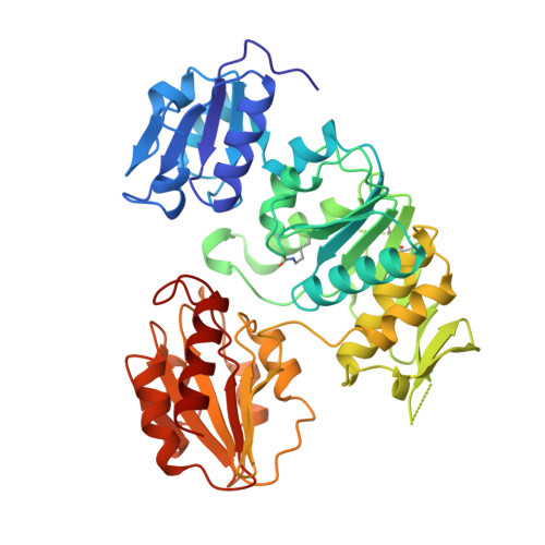
Crystal structure of UDP-N-acetylmuramoyl-L-alanine:D-glutamate ligase from Escherichia coli.

Bertrand, J.A.Auger, G.Fanchon, E.Martin, L.Blanot, D.van Heijenoort, J.Dideberg, O.

(1997) EMBO J 16: 3416-3425

  • DOI: https://doi.org/10.1093/emboj/16.12.3416
  • Primary Citation Related Structures: 
    1UAG

  • PubMed Abstract: 

    UDP-N-acetylmuramoyl-L-alanine:D-glutamate ligase (MurD) is a cytoplasmic enzyme involved in the biosynthesis of peptidoglycan which catalyzes the addition of D-glutamate to the nucleotide precursor UDP-N-acetylmuramoyl-L-alanine (UMA). The crystal structure of MurD in the presence of its substrate UMA has been solved to 1.9 A resolution. Phase information was obtained from multiple anomalous dispersion using the K-shell edge of selenium in combination with multiple isomorphous replacement. The structure comprises three domains of topology each reminiscent of nucleotide-binding folds: the N- and C-terminal domains are consistent with the dinucleotide-binding fold called the Rossmann fold, and the central domain with the mononucleotide-binding fold also observed in the GTPase family. The structure reveals the binding site of the substrate UMA, and comparison with known NTP complexes allows the identification of residues interacting with ATP. The study describes the first structure of the UDP-N-acetylmuramoyl-peptide ligase family.


  • Organizational Affiliation
    • Institut de Biologie Structurale Jean-Pierre Ebel (CEA-CNRS), Laboratoire de Cristallographie Macromoléculaire, Grenoble, France.

Macromolecule Content 

  • Total Structure Weight: 47.87 kDa 
  • Atom Count: 3,577 
  • Modeled Residue Count: 428 
  • Deposited Residue Count: 437 
  • Unique protein chains: 1

Macromolecules

Find similar proteins by:|  3D Structure
Entity ID: 1
MoleculeChains  Sequence LengthOrganismDetailsImage
UDP-N-ACETYLMURAMOYL-L-ALANINE/:D-GLUTAMATE LIGASE437Escherichia coliMutation(s): 1 
Gene Names: MURD GENE
EC: 6.3.2.9
UniProt
Find proteins for P14900 (Escherichia coli (strain K12))
Explore P14900 
Go to UniProtKB:  P14900
Entity Groups
Sequence Clusters30% Identity50% Identity70% Identity90% Identity95% Identity100% Identity
UniProt GroupP14900
Sequence Annotations
Expand
Reference Sequence

Small Molecules

Modified Residues  1 Unique
IDChains TypeFormula2D DiagramParent
KCX
Query on KCX
A
L-PEPTIDE LINKINGC7 H14 N2 O4LYS

Experimental Data & Validation

Experimental Data

  • Method: X-RAY DIFFRACTION
  • Resolution: 1.95 Å
  • R-Value Free:  0.235 (Depositor), 0.223 (DCC) 
  • R-Value Work:  0.190 (Depositor), 0.180 (DCC) 
  • R-Value Observed: 0.190 (Depositor) 
Space Group: P 41
Unit Cell:
Length ( Å )Angle ( ˚ )
a = 65.428α = 90
b = 65.428β = 90
c = 134.438γ = 90
Software Package:
Software NamePurpose
XDSdata scaling
MADSYSphasing
X-PLORmodel building
X-PLORrefinement
XDSdata reduction
X-PLORphasing

Structure Validation

View Full Validation Report


Ligand Structure Quality Assessment 


Entry History 

Deposition Data

Revision History  (Full details and data files)

  • Version 1.0: 1998-03-18
    Type: Initial release
  • Version 1.1: 2008-03-24
    Changes: Version format compliance
  • Version 1.2: 2011-07-13
    Changes: Version format compliance
  • Version 1.3: 2018-04-04
    Changes: Data collection
  • Version 1.4: 2018-04-11
    Changes: Data collection
  • Version 1.5: 2025-03-26
    Changes: Data collection, Database references, Derived calculations, Structure summary