1U4C | pdb_00001u4c

Structure of spindle checkpoint protein Bub3


Experimental Data Snapshot

  • Method: X-RAY DIFFRACTION
  • Resolution: 2.35 Å
  • R-Value Free: 
    0.288 (Depositor), 0.292 (DCC) 
  • R-Value Work: 
    0.196 (Depositor), 0.205 (DCC) 
  • R-Value Observed: 
    0.202 (Depositor) 

wwPDB Validation 3D Report Full Report

Validation slider image for 1U4C

This is version 1.5 of the entry. See complete history

Literature

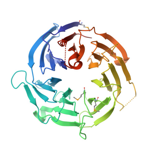
Crystal structure of the spindle assembly checkpoint protein Bub3

Larsen, N.A.Harrison, S.C.

(2004) J Mol Biology 344: 885-892

  • DOI: https://doi.org/10.1016/j.jmb.2004.09.094
  • Primary Citation Related Structures: 
    1U4C

  • PubMed Abstract: 

    Bub3 is one of at least six proteins that transmit the spindle assembly checkpoint signal. These proteins delay cell cycle progression from metaphase to anaphase in response to attachment defects between kinetochores and spindle microtubules and to tension defects between sister chromatids. To explore the molecular interactions mediated by Bub3, we have determined the crystal structure of the Saccharomyces cerevisiae protein Bub3p at 2.35 A resolution. Bub3p is a seven-blade beta-propeller, although its sequence diverges from that of other WD40 family members. Several loops are substantially elongated, but extra domains or insertions are not present at the termini. In particular, two extended loops project from the top face of the propeller, forming a cleft. Amino acid residues across the top face and one aspect of the lateral surface (spanning blades 5-6) are highly conserved among Bub3 proteins. We propose that these conserved surfaces are the loci for key interactions with conserved motifs in spindle checkpoint proteins Bub1 and Mad3/BubR1. Comparison of the Bub3 sequence to the WD40 protein, Rae1, shows high sequence conservation along the same surfaces. Rae1 interaction with Bub1 is, therefore, likely to involve a similar mode of binding.


  • Organizational Affiliation
    • Department of Biological Chemistry and Molecular Pharmacology, Harvard Medical School, Boston, MA, USA.

Macromolecule Content 

  • Total Structure Weight: 79.39 kDa 
  • Atom Count: 5,511 
  • Modeled Residue Count: 653 
  • Deposited Residue Count: 698 
  • Unique protein chains: 1

Macromolecules

Find similar proteins by:|  3D Structure
Entity ID: 1
MoleculeChains  Sequence LengthOrganismDetailsImage
Cell cycle arrest protein BUB3
A, B
349Saccharomyces cerevisiaeMutation(s): 3 
Gene Names: BUB3
UniProt
Find proteins for P26449 (Saccharomyces cerevisiae (strain ATCC 204508 / S288c))
Explore P26449 
Go to UniProtKB:  P26449
Entity Groups
Sequence Clusters30% Identity50% Identity70% Identity90% Identity95% Identity100% Identity
UniProt GroupP26449
Sequence Annotations
Expand
Reference Sequence

Small Molecules

Modified Residues  1 Unique
IDChains TypeFormula2D DiagramParent
MSE
Query on MSE
A, B
L-PEPTIDE LINKINGC5 H11 N O2 SeMET

Experimental Data & Validation

Experimental Data

  • Method: X-RAY DIFFRACTION
  • Resolution: 2.35 Å
  • R-Value Free:  0.288 (Depositor), 0.292 (DCC) 
  • R-Value Work:  0.196 (Depositor), 0.205 (DCC) 
  • R-Value Observed: 0.202 (Depositor) 
Space Group: P 32
Unit Cell:
Length ( Å )Angle ( ˚ )
a = 73.266α = 90
b = 73.266β = 90
c = 110.425γ = 120
Software Package:
Software NamePurpose
CNSrefinement
SCALEPACKdata scaling
SOLVEphasing
SHELXL-97refinement
HKL-2000data collection
HKL-2000data reduction

Structure Validation

View Full Validation Report


Entry History 

Deposition Data

Revision History  (Full details and data files)

  • Version 1.0: 2004-08-03
    Type: Initial release
  • Version 1.1: 2008-04-30
    Changes: Version format compliance
  • Version 1.2: 2011-07-13
    Changes: Version format compliance
  • Version 1.3: 2019-07-24
    Changes: Data collection, Derived calculations, Refinement description
  • Version 1.4: 2019-08-14
    Changes: Data collection
  • Version 1.5: 2024-11-06
    Changes: Data collection, Database references, Structure summary