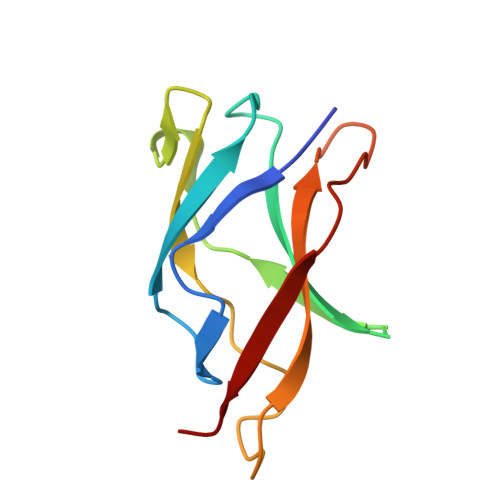
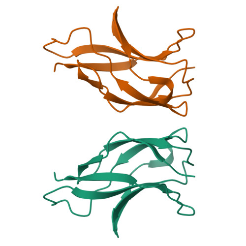
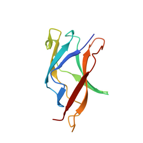
1U3J

Crystal structure of MLAV mutant of dimerisation domain of NF-kB p50 transcription factor


Experimental Data Snapshot

  • Method: X-RAY DIFFRACTION
  • Resolution: 1.90 Å
  • R-Value Free: 0.222 
  • R-Value Work: 0.182 
  • R-Value Observed: 0.184 

Starting Model: experimental
View more details

wwPDB Validation   3D Report Full Report


This is version 1.5 of the entry. See complete history


Literature

Snapshot of Protein Structure Evolution Reveals Conservation of Functional Dimerization through Intertwined Folding

Chirgadze, D.Y.Demydchuk, M.Becker, M.Moran, S.Paoli, M.

(2004) Structure 12: 1489-1494

  • DOI: https://doi.org/10.1016/j.str.2004.06.011
  • Primary Citation of Related Structures:  
    1U36, 1U3J, 1U3Y, 1U3Z, 1U41, 1U42

  • PubMed Abstract: 

    Protein-protein interactions govern a wide range of cellular processes. Molecular recognition responsible for homodimerization and heterodimerization in the rel/NF-kappaB family of eukaryotic transcription factors relies on a small cluster of hydrophobic residues. We have carried out a structural analysis of six NF-kappaB p50 dimer interface mutants; one of them revealed a remarkable alteration. One or possibly both its mutations cause a switch into an intertwined dimer, in which the molecular partners exchange nearly half of their fold. In spite of the extensive swapping of secondary structure elements, the topology within each counterpart is preserved, with a very similar overall structure and minimal changes at the interface. Thus intertwining rescues structure and function from a destabilizing mutation. Since the mutants originate from a directed evolution experiment and are functional, the data provide an evolutionary snapshot of how a protein structure can respond to mutations while maintaining a functional molecular architecture.


  • Organizational Affiliation

    Department of Biochemistry, University of Cambridge, Tennis Court Road, Cambridge, CB2 1GA, United Kingdom.


Macromolecules
Find similar proteins by:  (by identity cutoff)  |  3D Structure
Entity ID: 1
MoleculeChains Sequence LengthOrganismDetailsImage
Nuclear factor NF-kappa-B p105 subunit106Mus musculusMutation(s): 1 
Gene Names: Nfkb118033
UniProt & NIH Common Fund Data Resources
Find proteins for P25799 (Mus musculus)
Explore P25799 
Go to UniProtKB:  P25799
IMPC:  MGI:97312
Entity Groups  
Sequence Clusters30% Identity50% Identity70% Identity90% Identity95% Identity100% Identity
UniProt GroupP25799
Sequence Annotations
Expand
  • Reference Sequence
Experimental Data & Validation

Experimental Data

  • Method: X-RAY DIFFRACTION
  • Resolution: 1.90 Å
  • R-Value Free: 0.222 
  • R-Value Work: 0.182 
  • R-Value Observed: 0.184 
  • Space Group: P 41 21 2
Unit Cell:
Length ( Å )Angle ( ˚ )
a = 62.817α = 90
b = 62.817β = 90
c = 65.799γ = 90
Software Package:
Software NamePurpose
DENZOdata reduction
SCALEPACKdata scaling
AMoREphasing
REFMACrefinement

Structure Validation

View Full Validation Report


Entry History 

Deposition Data

Revision History  (Full details and data files)

  • Version 1.0: 2004-08-17
    Type: Initial release
  • Version 1.1: 2008-04-30
    Changes: Version format compliance
  • Version 1.2: 2011-07-13
    Changes: Version format compliance
  • Version 1.3: 2017-02-01
    Changes: Structure summary
  • Version 1.4: 2021-10-20
    Changes: Database references
  • Version 1.5: 2023-08-23
    Changes: Data collection, Refinement description