1U34 | pdb_00001u34

3D NMR structure of the first extracellular domain of CRFR-2beta, a type B1 G-protein coupled receptor


Experimental Data Snapshot

  • Method: SOLUTION NMR
  • Conformers Calculated: 100 
  • Conformers Submitted: 20 
  • Selection Criteria: structures with the lowest energy 

wwPDB Validation 3D Report Full Report

Validation slider image for 1U34

This is version 1.4 of the entry. See complete history

Literature

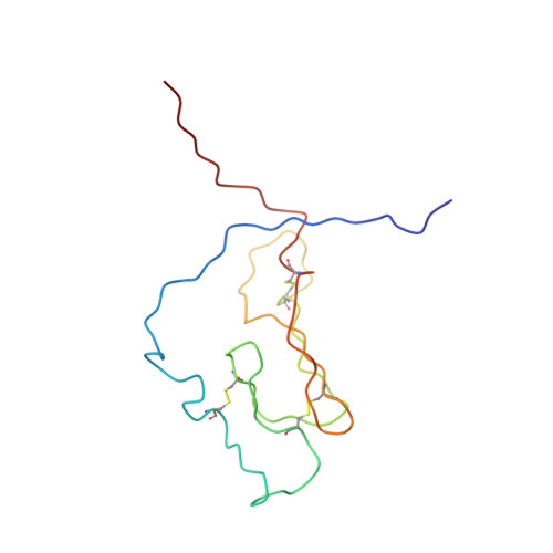
NMR structure and peptide hormone binding site of the first extracellular domain of a type B1 G protein-coupled receptor

Grace, C.R.Perrin, M.H.DiGruccio, M.R.Miller, C.L.Rivier, J.E.Vale, W.W.Riek, R.

(2004) Proc Natl Acad Sci U S A 101: 12836-12841

  • DOI: https://doi.org/10.1073/pnas.0404702101
  • Primary Citation Related Structures: 
    1U34

  • PubMed Abstract: 

    The corticotropin-releasing factor (CRF) ligand family has diverse effects on the CNS, including the modulation of the stress response. The ligands' effects are mediated by binding to CRF G protein-coupled receptors. We have determined the 3D NMR structure of the N-terminal extracellular domain (ECD1) of the mouse CRF receptor 2beta, which is the major ligand recognition domain, and identified its ligand binding site by chemical-shift perturbation experiments. The fold is identified as a short consensus repeat (SCR), a common protein interaction module. Mutagenesis reveals the integrity of the hormone-binding site in the full-length receptor. This study proposes that the ECD1 captures the C-terminal segment of the ligand, whose N terminus then penetrates into the transmembrane region of the receptor to initiate signaling. Key residues of SCR in the ECD1 are conserved in the G protein-coupled receptor subfamily, suggesting the SCR fold in all of the ECD1s of this subfamily.


  • Organizational Affiliation
    • Structural Biology Laboratory, and The Clayton Foundation Laboratories for Peptide Biology, The Salk Institute for Biological Studies, 10010 North Torrey Pines Road, La Jolla, CA 92037, USA. perrin@salk.edu

Macromolecule Content 

  • Total Structure Weight: 13.63 kDa 
  • Atom Count: 954 
  • Modeled Residue Count: 119 
  • Deposited Residue Count: 119 
  • Unique protein chains: 1

Macromolecules

Find similar proteins by:|  3D Structure
Entity ID: 1
MoleculeChains  Sequence LengthOrganismDetailsImage
Corticotropin releasing factor receptor 2119Mus musculusMutation(s): 0 
UniProt
Find proteins for Q60748 (Mus musculus)
Explore Q60748 
Go to UniProtKB:  Q60748
Entity Groups
Sequence Clusters30% Identity50% Identity70% Identity90% Identity95% Identity100% Identity
UniProt GroupQ60748
Sequence Annotations
Expand
Reference Sequence

Experimental Data & Validation

Experimental Data

  • Method: SOLUTION NMR
  • Conformers Calculated: 100 
  • Conformers Submitted: 20 
  • Selection Criteria: structures with the lowest energy 

Structure Validation

View Full Validation Report


Entry History 

Deposition Data

Revision History  (Full details and data files)

  • Version 1.0: 2004-09-07
    Type: Initial release
  • Version 1.1: 2008-04-30
    Changes: Version format compliance
  • Version 1.2: 2011-07-13
    Changes: Version format compliance
  • Version 1.3: 2022-03-02
    Changes: Database references, Derived calculations
  • Version 1.4: 2024-11-20
    Changes: Data collection, Structure summary