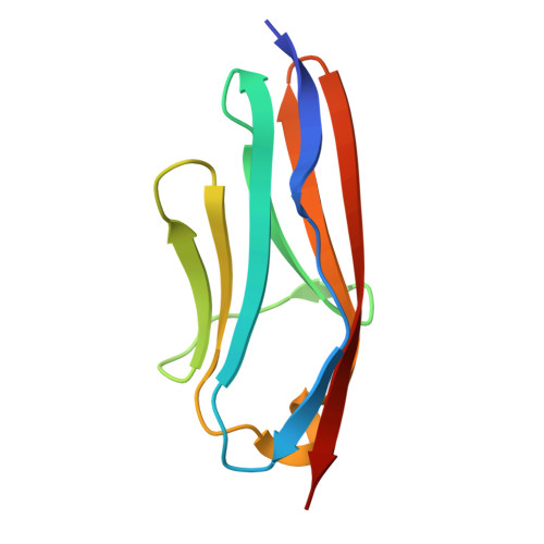
X-ray structure of engineered human Aortic Preferentially Expressed Protein-1 (APEG-1)
Manjasetty, B.A., Niesen, F.H., Scheich, C., Roske, Y., Gotz, F., Behlke, J., Sievert, V., Heinemann, U., Bussow, K.(2005) BMC Struct Biol 5: 21-21
- PubMed: 16354304
- DOI: https://doi.org/10.1186/1472-6807-5-21
- Primary Citation of Related Structures:
1U2H - PubMed Abstract:
Human Aortic Preferentially Expressed Protein-1 (APEG-1) is a novel specific smooth muscle differentiation marker thought to play a role in the growth and differentiation of arterial smooth muscle cells (SMCs). Good quality crystals that were suitable for X-ray crystallographic studies were obtained following the truncation of the 14 N-terminal amino acids of APEG-1, a region predicted to be disordered. The truncated protein (termed DeltaAPEG-1) consists of a single immunoglobulin (Ig) like domain which includes an Arg-Gly-Asp (RGD) adhesion recognition motif. The RGD motif is crucial for the interaction of extracellular proteins and plays a role in cell adhesion. The X-ray structure of DeltaAPEG-1 was determined and was refined to sub-atomic resolution (0.96 A). This is the best resolution for an immunoglobulin domain structure so far. The structure adopts a Greek-key beta-sandwich fold and belongs to the I (intermediate) set of the immunoglobulin superfamily. The residues lying between the beta-sheets form a hydrophobic core. The RGD motif folds into a 310 helix that is involved in the formation of a homodimer in the crystal which is mainly stabilized by salt bridges. Analytical ultracentrifugation studies revealed a moderate dissociation constant of 20 microM at physiological ionic strength, suggesting that APEG-1 dimerisation is only transient in the cell. The binding constant is strongly dependent on ionic strength. Our data suggests that the RGD motif might play a role not only in the adhesion of extracellular proteins but also in intracellular protein-protein interactions. However, it remains to be established whether the rather weak dimerisation of APEG-1 involving this motif is physiologically relevant.
Organizational Affiliation:
Protein Structure Factory, c/o BESSY GmbH, Albert-Einstein-Str. 15, 12489 Berlin, Germany. babu@bnl.gov


