1TZ6 | pdb_00001tz6

Crystal structure of aminoimidazole riboside kinase from Salmonella enterica complexed with aminoimidazole riboside and ATP analog


Experimental Data Snapshot

  • Method: X-RAY DIFFRACTION
  • Resolution: 2.70 Å
  • R-Value Free: 
    0.285 (Depositor), 0.285 (DCC) 
  • R-Value Work: 
    0.217 (Depositor), 0.217 (DCC) 
  • R-Value Observed: 
    0.217 (Depositor) 

Starting Model: experimental
View more details

wwPDB Validation 3D Report Full Report

Validation slider image for 1TZ6

Ligand Structure Quality Assessment 


This is version 1.3 of the entry. See complete history

Literature

Crystal Structure of an Aminoimidazole Riboside Kinase from Salmonella enterica; Implications for the Evolution of the Ribokinase Superfamily

Zhang, Y.Dougherty, M.Downs, D.M.Ealick, S.E.

(2004) Structure 12: 1809-1821

  • DOI: https://doi.org/10.1016/j.str.2004.07.020
  • Primary Citation Related Structures: 
    1TYY, 1TZ3, 1TZ6

  • PubMed Abstract: 

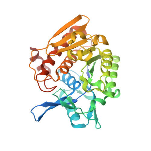
    The crystal structures of a Salmonella enterica aminoimidazole riboside (AIRs) kinase, its complex with the substrate AIRs, and its complex with AIRs and an ATP analog were determined at 2.6 angstroms, 2.9 angstroms, and 2.7 angstroms, respectively. The product of the Salmonella-specific gene stm4066, AIRs kinase, is a homodimer with one active site per monomer. The core structure, consisting of an eight-stranded beta sheet flanked by eight alpha helices, indicates that AIRs kinase is a member of the ribokinase superfamily. Unlike ribokinase and adenosine kinase in this superfamily, AIRs kinase does not show significant conformational changes upon substrate binding. The active site is covered by a lid formed by residues 16-28 and 86-100. A comparison of the structure of AIRs kinase with other ribokinase superfamily members suggests that the active site lid and conformational changes that occur upon substrate binding may be advanced features in the evolution of the ribokinase superfamily.


  • Organizational Affiliation
    • Department of Molecular Biology and Genetics, Cornell University, Ithaca, NY 14853, USA.

Macromolecule Content 

  • Total Structure Weight: 74.31 kDa 
  • Atom Count: 4,583 
  • Modeled Residue Count: 594 
  • Deposited Residue Count: 678 
  • Unique protein chains: 1

Macromolecules

Find similar proteins by:|  3D Structure
Entity ID: 1
MoleculeChains  Sequence LengthOrganismDetailsImage
putative sugar kinase
A, B
339Salmonella enterica subsp. enterica serovar Typhimurium str. LT2Mutation(s): 0 
Gene Names: Stm4066
EC: 2.7.1.4 (PDB Primary Data), 2.7.1.223 (UniProt)
UniProt
Find proteins for Q8ZKR2 (Salmonella typhimurium (strain LT2 / SGSC1412 / ATCC 700720))
Explore Q8ZKR2 
Go to UniProtKB:  Q8ZKR2
Entity Groups
Sequence Clusters30% Identity50% Identity70% Identity90% Identity95% Identity100% Identity
UniProt GroupQ8ZKR2
Sequence Annotations
Expand
Reference Sequence

Small Molecules

Ligands 4 Unique
IDChains Name / Formula / InChI Key2D Diagram3D Interactions
ACP

Query on ACP


Download:Ideal Coordinates CCD File
F [auth A],
K [auth B]
PHOSPHOMETHYLPHOSPHONIC ACID ADENYLATE ESTER
C11 H18 N5 O12 P3
UFZTZBNSLXELAL-IOSLPCCCSA-N
AIS

Query on AIS


Download:Ideal Coordinates CCD File
G [auth A]5-AMINOIMIDAZOLE RIBONUCLEOSIDE
C8 H13 N3 O4
NKYAAYKKNSYIIW-XVFCMESISA-N
K

Query on K


Download:Ideal Coordinates CCD File
D [auth A],
E [auth A],
I [auth B],
J [auth B]
POTASSIUM ION
K
NPYPAHLBTDXSSS-UHFFFAOYSA-N
MG

Query on MG


Download:Ideal Coordinates CCD File
C [auth A],
H [auth B]
MAGNESIUM ION
Mg
JLVVSXFLKOJNIY-UHFFFAOYSA-N

Experimental Data & Validation

Experimental Data

  • Method: X-RAY DIFFRACTION
  • Resolution: 2.70 Å
  • R-Value Free:  0.285 (Depositor), 0.285 (DCC) 
  • R-Value Work:  0.217 (Depositor), 0.217 (DCC) 
  • R-Value Observed: 0.217 (Depositor) 
Space Group: P 21 21 21
Unit Cell:
Length ( Å )Angle ( ˚ )
a = 137.007α = 90
b = 54.101β = 90
c = 89.895γ = 90
Software Package:
Software NamePurpose
CNSrefinement
HKL-2000data reduction
SCALEPACKdata scaling
CNSphasing

Structure Validation

View Full Validation Report


Ligand Structure Quality Assessment 


Entry History 

Deposition Data

Revision History  (Full details and data files)

  • Version 1.0: 2004-10-12
    Type: Initial release
  • Version 1.1: 2008-04-30
    Changes: Version format compliance
  • Version 1.2: 2011-07-13
    Changes: Version format compliance
  • Version 1.3: 2023-08-23
    Changes: Data collection, Database references, Derived calculations, Refinement description