1TWW

Dihydropteroate Synthetase, With Bound Substrate Analogue PtPP, From Bacillus anthracis


Experimental Data Snapshot

  • Method: X-RAY DIFFRACTION
  • Resolution: 2.50 Å
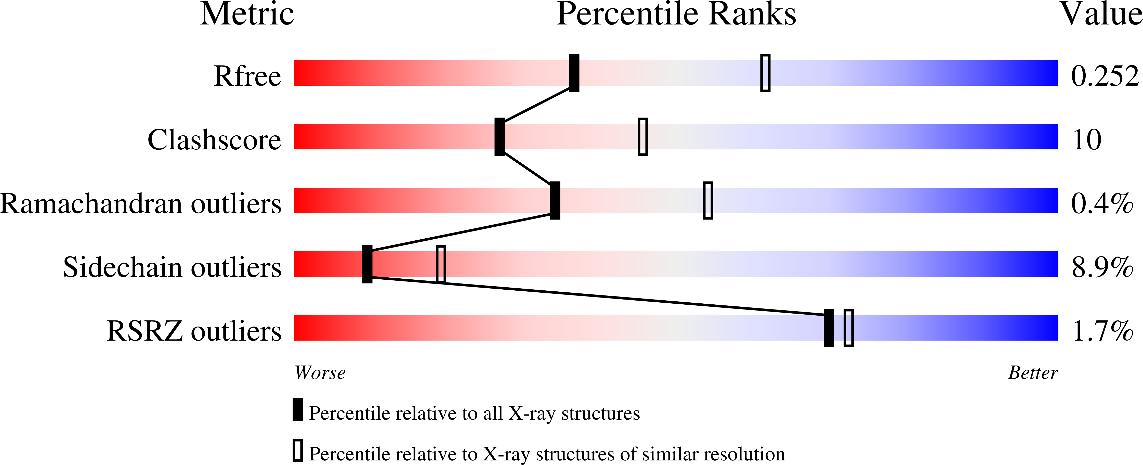
  • R-Value Free: 0.254 
  • R-Value Work: 0.211 
  • R-Value Observed: 0.213 

Starting Model: experimental
View more details

wwPDB Validation   3D Report Full Report


Ligand Structure Quality Assessment 


This is version 1.4 of the entry. See complete history


Literature

Crystal Structure of 7,8-Dihydropteroate Synthase from Bacillus anthracis; Mechanism and Novel Inhibitor Design.

Babaoglu, K.Qi, J.Lee, R.E.White, S.W.

(2004) Structure 12: 1705-1717

  • DOI: https://doi.org/10.1016/j.str.2004.07.011
  • Primary Citation of Related Structures:  
    1TWS, 1TWW, 1TWZ, 1TX0, 1TX2

  • PubMed Abstract: 

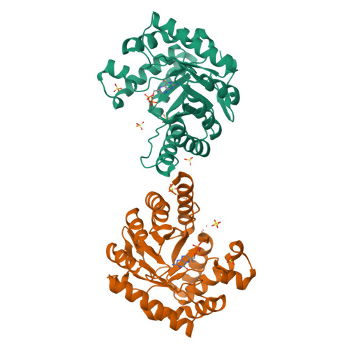
    Dihydropterate synthase (DHPS) is the target for the sulfonamide class of antibiotics, but increasing resistance has encouraged the development of new therapeutic agents against this enzyme. One approach is to identify molecules that occupy the pterin binding pocket which is distinct from the pABA binding pocket that binds sulfonamides. Toward this goal, we present five crystal structures of DHPS from Bacillus anthracis, a well-documented bioterrorism agent. Three DHPS structures are already known, but our B. anthracis structures provide new insights into the enzyme mechanism. We show how an arginine side chain mimics the pterin ring in binding within the pterin binding pocket. The structures of two substrate analog complexes and the first structure of a DHPS-product complex offer new insights into the catalytic mechanism and the architecture of the pABA binding pocket. Finally, as an initial step in the development of pterin-based inhibitors, we present the structure of DHPS complexed with 5-nitro-6-methylamino-isocytosine.


  • Organizational Affiliation

    Department of Structural Biology, St Jude Children's Research Hospital, Memphis, TN 38105, USA.


Macromolecules
Find similar proteins by:  (by identity cutoff)  |  3D Structure
Entity ID: 1
MoleculeChains Sequence LengthOrganismDetailsImage
DHPS, Dihydropteroate synthase
A, B
297Bacillus anthracis str. A2012Mutation(s): 0 
Gene Names: folp
EC: 2.5.1.15
UniProt
Find proteins for Q81VW8 (Bacillus anthracis)
Explore Q81VW8 
Go to UniProtKB:  Q81VW8
Entity Groups  
Sequence Clusters30% Identity50% Identity70% Identity90% Identity95% Identity100% Identity
UniProt GroupQ81VW8
Sequence Annotations
Expand
  • Reference Sequence
Small Molecules
Ligands 2 Unique
IDChains Name / Formula / InChI Key2D Diagram3D Interactions
HH2
Query on HH2

Download Ideal Coordinates CCD File 
G [auth A],
L [auth B]
6-HYDROXYMETHYLPTERIN-DIPHOSPHATE
C7 H9 N5 O8 P2
AMDUVUKDRBIVAH-UHFFFAOYSA-N
SO4
Query on SO4

Download Ideal Coordinates CCD File 
C [auth A]
D [auth A]
E [auth A]
F [auth A]
H [auth B]
C [auth A],
D [auth A],
E [auth A],
F [auth A],
H [auth B],
I [auth B],
J [auth B],
K [auth B]
SULFATE ION
O4 S
QAOWNCQODCNURD-UHFFFAOYSA-L
Experimental Data & Validation

Experimental Data

  • Method: X-RAY DIFFRACTION
  • Resolution: 2.50 Å
  • R-Value Free: 0.254 
  • R-Value Work: 0.211 
  • R-Value Observed: 0.213 
  • Space Group: P 62 2 2
Unit Cell:
Length ( Å )Angle ( ˚ )
a = 98.239α = 90
b = 98.239β = 90
c = 262.61γ = 120
Software Package:
Software NamePurpose
REFMACrefinement
MAR345data collection
SCALEPACKdata scaling
CNSphasing

Structure Validation

View Full Validation Report


Ligand Structure Quality Assessment 


Entry History 

Deposition Data

Revision History  (Full details and data files)

  • Version 1.0: 2004-09-14
    Type: Initial release
  • Version 1.1: 2008-04-30
    Changes: Version format compliance
  • Version 1.2: 2011-07-13
    Changes: Advisory, Derived calculations, Refinement description, Source and taxonomy, Version format compliance
  • Version 1.3: 2017-10-11
    Changes: Refinement description
  • Version 1.4: 2023-08-23
    Changes: Data collection, Database references, Derived calculations, Refinement description