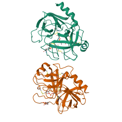
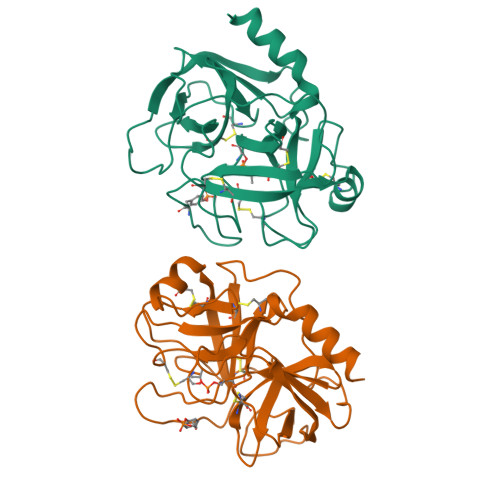
Crystal structure of human trypsin 1: unexpected phosphorylation of Tyr151.
Gaboriaud, C., Serre, L., Guy-Crotte, O., Forest, E., Fontecilla-Camps, J.C.(1996) J Mol Biology 259: 995-1010
- PubMed: 8683601
- DOI: https://doi.org/10.1006/jmbi.1996.0376
- Primary Citation of Related Structures:
1TRN - PubMed Abstract:
The X-ray structure of human trypsin 1 has been determined in the presence of diisopropyl-phosphofluoridate by the molecular replacement method and refined at a resolution of 2.2 A to an R-factor of 18%. Crystals belong to the space group P4, with two independent molecules in the asymmetric unit packing as crystallographic tetramers. This study was performed in order to seek possible structural peculiarities of human trypsin 1, suggested by some striking differences in its biochemical behavior as compared to other trypsins of mammalian species. Its fold is, in fact, very similar to those of the bovine, rat and porcine trypsins, with root-mean-square differences in the 0.4 to 0.6 A range for all 223 C alpha positions. The most unexpected feature of the human trypsin 1 structure is in the phosphorylated state of tyrosine residue 151 in the present X-ray study. This feature was confirmed by mass spectrometry on the same inhibited sample and also on the native enzyme. This phosphorylation strengthens the outstanding clustering of highly negative or highly positive electrostatic surface potentials. The peculiar inhibitory behaviour of pancreatic secretory trypsin inhibitors of the Kazal type on this enzyme is discussed as a possible consequence of these properties. A charged surface loop has also been interpreted as an epitope site recognised by a monoclonal antibody specific to human trypsin 1.
Organizational Affiliation:
Laboratoire de Cristallographie et Cristallogénèse des Protéines, Institut de Biologie Structurale Jean-Pierre Ebel (CEA-CNRS), Grenoble, France.


