1TRI | pdb_00001tri

THE CRYSTAL STRUCTURE OF AN ENGINEERED MONOMERIC TRIOSEPHOSPHATE ISOMERASE, MONOTIM: THE CORRECT MODELLING OF AN EIGHT-RESIDUE LOOP


Experimental Data Snapshot

  • Method: X-RAY DIFFRACTION
  • Resolution: 2.40 Å
  • R-Value Work: 
    0.165 (Depositor) 
  • R-Value Observed: 
    0.165 (Depositor) 

wwPDB Validation 3D Report Full Report

Validation slider image for 1TRI

This is version 1.6 of the entry. See complete history

Literature

The crystal structure of an engineered monomeric triosephosphate isomerase, monoTIM: the correct modelling of an eight-residue loop.

Borchert, T.V.Abagyan, R.Kishan, K.V.Zeelen, J.P.Wierenga, R.K.

(1993) Structure 1: 205-213

  • DOI: https://doi.org/10.1016/0969-2126(93)90021-8
  • Primary Citation Related Structures: 
    1TRI

  • PubMed Abstract: 

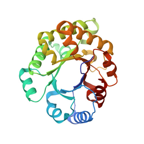
    The triosephosphate isomerase (TIM) fold is found in several different classes of enzymes, most of which are oligomers; TIM itself always functions as a very tight dimer. It has recently been shown that a monomeric form of TIM ('monoTIM') can be constructed by replacing a 15-residue interface loop, loop-3, with an eight-residue fragment; modelling suggests that this should result in a short strain-free turn, resulting in the subsequent helix, helix-A3, having an additional turn at its amino terminus. The crystal structure of monoTIM shows that it retains the characteristic TIM-barrel (betaalpha)8-fold and that the new loop has a structure very close to that predicted. Two other interface loops, loop-1 and loop-4, which contain the active site residues Lys13 and His95, respectively, show significant changes in structure in monoTIM compared with dimeric wild-type TIM. The observed structural differences between monoTIM and wild-type TIM indicate that the dimeric appearance of TIM determines the location and conformation of two of the four catalytic residues.


  • Organizational Affiliation
    • European Molecular Biology Laboratory, Meyerhofstrasse 1, Postfach 102209, D69012 Heidelberg, Germany.

Macromolecule Content 

  • Total Structure Weight: 26.19 kDa 
  • Atom Count: 1,831 
  • Modeled Residue Count: 239 
  • Deposited Residue Count: 243 
  • Unique protein chains: 1

Macromolecules

Find similar proteins by:|  3D Structure
Entity ID: 1
MoleculeChains  Sequence LengthOrganismDetailsImage
TRIOSEPHOSPHATE ISOMERASE243Trypanosoma brucei bruceiMutation(s): 7 
EC: 5.3.1.1
UniProt
Find proteins for P04789 (Trypanosoma brucei brucei)
Explore P04789 
Go to UniProtKB:  P04789
Entity Groups
Sequence Clusters30% Identity50% Identity70% Identity90% Identity95% Identity100% Identity
UniProt GroupP04789
Sequence Annotations
Expand
Reference Sequence

Small Molecules

Ligands 1 Unique
IDChains Name / Formula / InChI Key2D Diagram3D Interactions
SO4

Query on SO4


Download:Ideal Coordinates CCD File
B [auth A]SULFATE ION
O4 S
QAOWNCQODCNURD-UHFFFAOYSA-L

Experimental Data & Validation

Experimental Data

  • Method: X-RAY DIFFRACTION
  • Resolution: 2.40 Å
  • R-Value Work:  0.165 (Depositor) 
  • R-Value Observed: 0.165 (Depositor) 
Space Group: C 1 2 1
Unit Cell:
Length ( Å )Angle ( ˚ )
a = 83.78α = 90
b = 42.8β = 108.22
c = 68.84γ = 90
Software Package:
Software NamePurpose
X-PLORmodel building
TNTrefinement
X-PLORrefinement
X-PLORphasing

Structure Validation

View Full Validation Report


Entry History 

Deposition Data

Revision History  (Full details and data files)

  • Version 1.0: 1994-07-31
    Type: Initial release
  • Version 1.1: 2008-03-24
    Changes: Version format compliance
  • Version 1.2: 2011-07-13
    Changes: Version format compliance
  • Version 1.3: 2019-07-17
    Changes: Data collection, Other, Refinement description
  • Version 1.4: 2019-08-14
    Changes: Data collection, Refinement description
  • Version 1.5: 2021-11-03
    Changes: Database references, Derived calculations
  • Version 1.6: 2024-02-14
    Changes: Data collection