1TPH | pdb_00001tph

1.8 ANGSTROMS CRYSTAL STRUCTURE OF WILD TYPE CHICKEN TRIOSEPHOSPHATE ISOMERASE-PHOSPHOGLYCOLOHYDROXAMATE COMPLEX


Experimental Data Snapshot

  • Method: X-RAY DIFFRACTION
  • Resolution: 1.80 Å
  • R-Value Observed: 
    0.185 (Depositor) 

wwPDB Validation 3D Report Full Report

Validation slider image for 1TPH

This is version 1.3 of the entry. See complete history

Literature

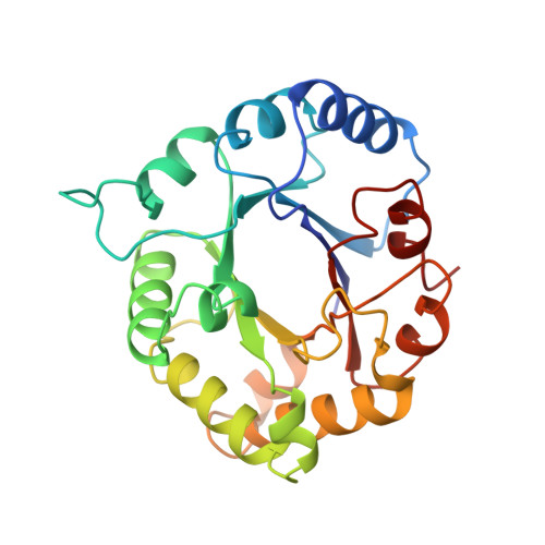
Crystal structure of recombinant chicken triosephosphate isomerase-phosphoglycolohydroxamate complex at 1.8-A resolution.

Zhang, Z.Sugio, S.Komives, E.A.Liu, K.D.Knowles, J.R.Petsko, G.A.Ringe, D.

(1994) Biochemistry 33: 2830-2837

  • DOI: https://doi.org/10.1021/bi00176a012
  • Primary Citation Related Structures: 
    1TPH

  • PubMed Abstract: 

    The crystal structure of recombinant chicken triosephosphate isomerase (TIM, E.C. 5.3.1.1) complexed with the intermediate analogue phosphoglycolohydroxamate (PGH) has been solved by the method of molecular replacement and refined to an R-factor of 18.5% at 1.8-A resolution. The structure is essentially identical to that of the yeast TIM-PGH complex [Davenport, R. C., et al. (1991) Biochemistry 30, 5821-5826] determined earlier and refined at comparable resolution. This identity extends to the high-energy conformations of the active-site residues Lys13 and Ser211, as well as the positions of several bound water molecules that are retained in the active site when PGH is bound. Comparison with the structure of uncomplexed chicken TIM shows that the catalytic base, Glu165, moves several angstroms when PGH binds. This movement may provide a trigger for a larger conformational change, one of 7 A, in a loop near the active site, which folds down like a lid to shield the bound inhibitor and catalytic residues from contact with bulk solvent. These same conformational changes were seen in crystalline yeast TIM upon binding of PGH; their occurrence here in a different crystal form of TIM eliminates the possibility that they are an artifact of crystal packing.


  • Organizational Affiliation
    • Department of Biochemistry, Rosenstiel Basic Medical Sciences Research Center, Brandeis University, Waltham, Massachusetts 02254-9110.

Macromolecule Content 

  • Total Structure Weight: 53.42 kDa 
  • Atom Count: 3,981 
  • Modeled Residue Count: 490 
  • Deposited Residue Count: 494 
  • Unique protein chains: 1

Macromolecules

Find similar proteins by:|  3D Structure
Entity ID: 1
MoleculeChains  Sequence LengthOrganismDetailsImage
TRIOSEPHOSPHATE ISOMERASEA [auth 1],
B [auth 2]
247Gallus gallusMutation(s): 0 
EC: 5.3.1.1 (PDB Primary Data), 4.2.3.3 (UniProt)
UniProt
Find proteins for P00940 (Gallus gallus)
Explore P00940 
Go to UniProtKB:  P00940
Entity Groups
Sequence Clusters30% Identity50% Identity70% Identity90% Identity95% Identity100% Identity
UniProt GroupP00940
Sequence Annotations
Expand
Reference Sequence

Small Molecules

Ligands 1 Unique
IDChains Name / Formula / InChI Key2D Diagram3D Interactions
PGH

Query on PGH


Download:Ideal Coordinates CCD File
C [auth 1],
D [auth 2]
PHOSPHOGLYCOLOHYDROXAMIC ACID
C2 H6 N O6 P
BAXHHWZKQZIJID-UHFFFAOYSA-N

Experimental Data & Validation

Experimental Data

  • Method: X-RAY DIFFRACTION
  • Resolution: 1.80 Å
  • R-Value Observed: 0.185 (Depositor) 
Space Group: P 21 21 21
Unit Cell:
Length ( Å )Angle ( ˚ )
a = 136.4α = 90
b = 74β = 90
c = 57.2γ = 90
Software Package:
Software NamePurpose
PROLSQrefinement

Structure Validation

View Full Validation Report


Entry History 

Deposition Data

Revision History  (Full details and data files)

  • Version 1.0: 1994-04-30
    Type: Initial release
  • Version 1.1: 2008-03-03
    Changes: Version format compliance
  • Version 1.2: 2011-07-13
    Changes: Version format compliance
  • Version 1.3: 2024-02-14
    Changes: Data collection, Database references, Derived calculations, Other