1TLL | pdb_00001tll

CRYSTAL STRUCTURE OF RAT NEURONAL NITRIC-OXIDE SYNTHASE REDUCTASE MODULE AT 2.3 A RESOLUTION.


Experimental Data Snapshot

  • Method: X-RAY DIFFRACTION
  • Resolution: 2.30 Å
  • R-Value Free: 
    0.272 (Depositor), 0.261 (DCC) 
  • R-Value Work: 
    0.244 (Depositor), 0.236 (DCC) 
  • R-Value Observed: 
    0.244 (Depositor) 

Starting Model: experimental
View more details

wwPDB Validation 3D Report Full Report

Validation slider image for 1TLL

Ligand Structure Quality Assessment 


This is version 1.3 of the entry. See complete history

Literature

Structural basis for isozyme-specific regulation of electron transfer in nitric-oxide synthase

Garcin, E.D.Bruns, C.M.Lloyd, S.J.Hosfield, D.J.Tiso, M.Gachhui, R.Stuehr, D.J.Tainer, J.A.Getzoff, E.D.

(2004) J Biological Chem 279: 37918-37927

  • DOI: https://doi.org/10.1074/jbc.M406204200
  • Primary Citation Related Structures: 
    1TLL

  • PubMed Abstract: 

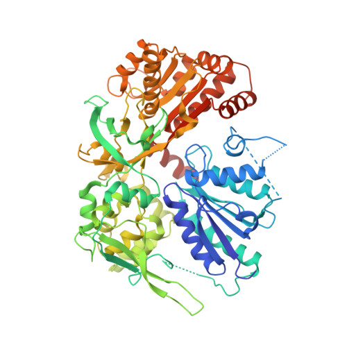
    Three nitric-oxide synthase (NOS) isozymes play crucial, but distinct, roles in neurotransmission, vascular homeostasis, and host defense, by catalyzing Ca(2+)/calmodulin-triggered NO synthesis. Here, we address current questions regarding NOS activity and regulation by combining mutagenesis and biochemistry with crystal structure determination of a fully assembled, electron-supplying, neuronal NOS reductase dimer. By integrating these results, we structurally elucidate the unique mechanisms for isozyme-specific regulation of electron transfer in NOS. Our discovery of the autoinhibitory helix, its placement between domains, and striking similarities with canonical calmodulin-binding motifs, support new mechanisms for NOS inhibition. NADPH, isozyme-specific residue Arg(1400), and the C-terminal tail synergistically repress NOS activity by locking the FMN binding domain in an electron-accepting position. Our analyses suggest that calmodulin binding or C-terminal tail phosphorylation frees a large scale swinging motion of the entire FMN domain to deliver electrons to the catalytic module in the holoenzyme.


  • Organizational Affiliation
    • Department of Molecular Biology and the Skaggs Institute for Chemical Biology, The Scripps Research Institute, La Jolla, California 92037, USA. edg@scripps.edu

Macromolecule Content 

  • Total Structure Weight: 160.15 kDa 
  • Atom Count: 10,408 
  • Modeled Residue Count: 1,246 
  • Deposited Residue Count: 1,376 
  • Unique protein chains: 1

Macromolecules

Find similar proteins by:|  3D Structure
Entity ID: 1
MoleculeChains  Sequence LengthOrganismDetailsImage
Nitric-oxide synthase, brain
A, B
688Rattus norvegicusMutation(s): 0 
Gene Names: NOS1BNOS
EC: 1.14.13.39
UniProt
Find proteins for P29476 (Rattus norvegicus)
Explore P29476 
Go to UniProtKB:  P29476
Entity Groups
Sequence Clusters30% Identity50% Identity70% Identity90% Identity95% Identity100% Identity
UniProt GroupP29476
Sequence Annotations
Expand
Reference Sequence

Small Molecules

Ligands 4 Unique
IDChains Name / Formula / InChI Key2D Diagram3D Interactions
FAD

Query on FAD


Download:Ideal Coordinates CCD File
E [auth A],
I [auth B]
FLAVIN-ADENINE DINUCLEOTIDE
C27 H33 N9 O15 P2
VWWQXMAJTJZDQX-UYBVJOGSSA-N
NAP

Query on NAP


Download:Ideal Coordinates CCD File
F [auth A],
J [auth B]
NADP NICOTINAMIDE-ADENINE-DINUCLEOTIDE PHOSPHATE
C21 H28 N7 O17 P3
XJLXINKUBYWONI-NNYOXOHSSA-N
FMN

Query on FMN


Download:Ideal Coordinates CCD File
D [auth A],
H [auth B]
FLAVIN MONONUCLEOTIDE
C17 H21 N4 O9 P
FVTCRASFADXXNN-SCRDCRAPSA-N
SO3

Query on SO3


Download:Ideal Coordinates CCD File
C [auth A],
G [auth B]
SULFITE ION
O3 S
LSNNMFCWUKXFEE-UHFFFAOYSA-L

Experimental Data & Validation

Experimental Data

  • Method: X-RAY DIFFRACTION
  • Resolution: 2.30 Å
  • R-Value Free:  0.272 (Depositor), 0.261 (DCC) 
  • R-Value Work:  0.244 (Depositor), 0.236 (DCC) 
  • R-Value Observed: 0.244 (Depositor) 
Space Group: P 1
Unit Cell:
Length ( Å )Angle ( ˚ )
a = 65.756α = 76.8
b = 69.174β = 72.07
c = 82.628γ = 67.14
Software Package:
Software NamePurpose
CNSrefinement
DENZOdata reduction
SCALEPACKdata scaling
AMoREphasing

Structure Validation

View Full Validation Report


Ligand Structure Quality Assessment 


Entry History 

Deposition Data

Revision History  (Full details and data files)

  • Version 1.0: 2004-08-31
    Type: Initial release
  • Version 1.1: 2008-04-30
    Changes: Version format compliance
  • Version 1.2: 2011-07-13
    Changes: Version format compliance
  • Version 1.3: 2023-08-23
    Changes: Data collection, Database references, Derived calculations, Refinement description