1TLJ | pdb_00001tlj

Crystal Structure of Conserved Protein of Unknown Function SSO0622 from Sulfolobus solfataricus


Experimental Data Snapshot

  • Method: X-RAY DIFFRACTION
  • Resolution: 2.80 Å
  • R-Value Free: 
    0.272 (Depositor), 0.235 (DCC) 
  • R-Value Work: 
    0.223 (Depositor), 0.239 (DCC) 
  • R-Value Observed: 
    0.249 (Depositor) 

wwPDB Validation 3D Report Full Report

Validation slider image for 1TLJ

This is version 1.4 of the entry. See complete history

Literature

Structural and functional characterization of the TYW3/Taw3 class of SAM-dependent methyltransferases.

Currie, M.A.Brown, G.Wong, A.Ohira, T.Sugiyama, K.Suzuki, T.Yakunin, A.F.Jia, Z.

(2017) RNA 23: 346-354

  • DOI: https://doi.org/10.1261/rna.057943.116
  • Primary Citation Related Structures: 
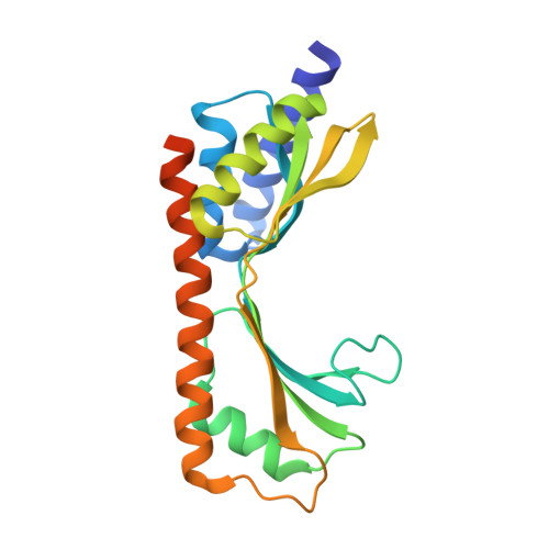
    1TLJ

  • PubMed Abstract: 

    S -adenosylmethionine (SAM)-dependent methyltransferases regulate a wide range of biological processes through the modification of proteins, nucleic acids, polysaccharides, as well as various metabolites. TYW3/Taw3 is a SAM-dependent methyltransferase responsible for the formation of a tRNA modification known as wybutosine and its derivatives that are required for accurate decoding in protein synthesis. Here, we report the crystal structure of Taw3, a homolog of TYW3 from Sulfolobus solfataricus , which revealed a novel α/β fold. The sequence motif (S/T)xSSCxGR and invariant aspartate and histidine, conserved in TYW3/Taw3, cluster to form the catalytic center. These structural and sequence features indicate that TYW3/Taw3 proteins constitute a distinct class of SAM-dependent methyltransferases. Using site-directed mutagenesis along with in vivo complementation assays combined with mass spectrometry as well as ligand docking and cofactor binding assays, we have identified the active site of TYW3 and residues essential for cofactor binding and methyltransferase activity.


  • Organizational Affiliation
    • Department of Biomedical and Molecular Sciences, Queen's University, Kingston, Ontario K7L 3N6, Canada.

Macromolecule Content 

  • Total Structure Weight: 49.27 kDa 
  • Atom Count: 3,346 
  • Modeled Residue Count: 377 
  • Deposited Residue Count: 426 
  • Unique protein chains: 1

Macromolecules

Find similar proteins by:|  3D Structure
Entity ID: 1
MoleculeChains  Sequence LengthOrganismDetailsImage
Hypothetical UPF0130 protein SSO0622
A, B
213Saccharolobus solfataricusMutation(s): 0 
Gene Names: SSO0622C08_028
EC: 2.1.1.282
UniProt
Find proteins for Q9UX16 (Saccharolobus solfataricus (strain ATCC 35092 / DSM 1617 / JCM 11322 / P2))
Explore Q9UX16 
Go to UniProtKB:  Q9UX16
Entity Groups
Sequence Clusters30% Identity50% Identity70% Identity90% Identity95% Identity100% Identity
UniProt GroupQ9UX16
Sequence Annotations
Expand
Reference Sequence

Experimental Data & Validation

Experimental Data

  • Method: X-RAY DIFFRACTION
  • Resolution: 2.80 Å
  • R-Value Free:  0.272 (Depositor), 0.235 (DCC) 
  • R-Value Work:  0.223 (Depositor), 0.239 (DCC) 
  • R-Value Observed: 0.249 (Depositor) 
Space Group: P 32 2 1
Unit Cell:
Length ( Å )Angle ( ˚ )
a = 92.114α = 90
b = 92.114β = 90
c = 110.907γ = 120
Software Package:
Software NamePurpose
HKL-2000data collection
SCALEPACKdata scaling
SOLVEphasing
CNSrefinement
HKL-2000data reduction

Structure Validation

View Full Validation Report


Entry History 

Revision History  (Full details and data files)

  • Version 1.0: 2004-08-03
    Type: Initial release
  • Version 1.1: 2008-04-30
    Changes: Version format compliance
  • Version 1.2: 2011-07-13
    Changes: Version format compliance
  • Version 1.3: 2020-07-29
    Changes: Database references, Derived calculations
  • Version 1.4: 2024-02-14
    Changes: Data collection, Database references