1TK5 | pdb_00001tk5

T7 DNA polymerase binary complex with 8 oxo guanosine in the templating strand


Experimental Data Snapshot

  • Method: X-RAY DIFFRACTION
  • Resolution: 2.20 Å
  • R-Value Free: 
    0.246 (Depositor), 0.241 (DCC) 
  • R-Value Work: 
    0.214 (Depositor), 0.209 (DCC) 
  • R-Value Observed: 
    0.214 (Depositor) 

Starting Model: experimental
View more details

wwPDB Validation 3D Report Full Report

Validation slider image for 1TK5

This is version 1.4 of the entry. See complete history

Literature

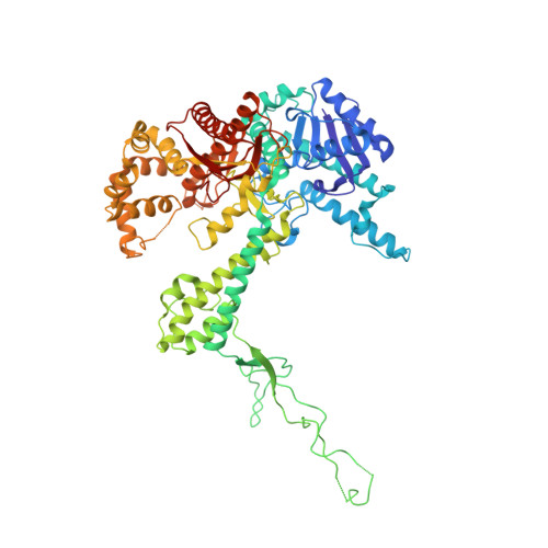
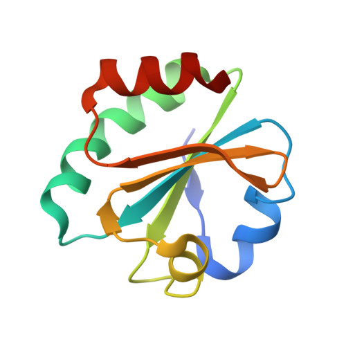
Structural basis for the dual coding potential of 8-oxoguanosine by a high-fidelity DNA polymerase.

Brieba, L.G.Eichman, B.F.Kokoska, R.J.Doublie, S.Kunkel, T.A.Ellenberger, T.

(2004) EMBO J 23: 3452-3461

  • DOI: https://doi.org/10.1038/sj.emboj.7600354
  • Primary Citation Related Structures: 
    1T8E, 1TK0, 1TK5, 1TK8, 1TKD

  • PubMed Abstract: 

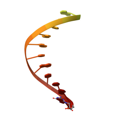
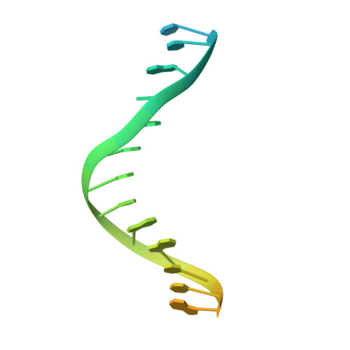
    Accurate DNA replication involves polymerases with high nucleotide selectivity and proofreading activity. We show here why both fidelity mechanisms fail when normally accurate T7 DNA polymerase bypasses the common oxidative lesion 8-oxo-7, 8-dihydro-2'-deoxyguanosine (8oG). The crystal structure of the polymerase with 8oG templating dC insertion shows that the O8 oxygen is tolerated by strong kinking of the DNA template. A model of a corresponding structure with dATP predicts steric and electrostatic clashes that would reduce but not eliminate insertion of dA. The structure of a postinsertional complex shows 8oG(syn).dA (anti) in a Hoogsteen-like base pair at the 3' terminus, and polymerase interactions with the minor groove surface of the mismatch that mimic those with undamaged, matched base pairs. This explains why translesion synthesis is permitted without proofreading of an 8oG.dA mismatch, thus providing insight into the high mutagenic potential of 8oG.


  • Organizational Affiliation
    • Department of Biological Chemistry and Molecular Pharmacology, Harvard Medical School, Boston, MA 02115, USA.

Macromolecule Content 

  • Total Structure Weight: 106.13 kDa 
  • Atom Count: 7,441 
  • Modeled Residue Count: 810 
  • Deposited Residue Count: 854 
  • Unique protein chains: 2
  • Unique nucleic acid chains: 2

Macromolecules


Find similar proteins by:|  3D Structure
Entity ID: 3
MoleculeChains  Sequence LengthOrganismDetailsImage
DNA polymeraseC [auth A]698Escherichia phage T7Mutation(s): 0 
Gene Names: 5
EC: 2.7.7.7 (PDB Primary Data), 3.1.11 (UniProt)
UniProt
Find proteins for P00581 (Escherichia phage T7)
Explore P00581 
Go to UniProtKB:  P00581
Entity Groups
Sequence Clusters30% Identity50% Identity70% Identity90% Identity95% Identity100% Identity
UniProt GroupP00581
Sequence Annotations
Expand
Reference Sequence
Find similar proteins by:|  3D Structure
Entity ID: 4
MoleculeChains  Sequence LengthOrganismDetailsImage
Thioredoxin 1D [auth B]108Escherichia coliMutation(s): 0 
Gene Names: 
UniProt
Find proteins for P0AA25 (Escherichia coli (strain K12))
Explore P0AA25 
Go to UniProtKB:  P0AA25
Entity Groups
Sequence Clusters30% Identity50% Identity70% Identity90% Identity95% Identity100% Identity
UniProt GroupP0AA25
Sequence Annotations
Expand
Reference Sequence
Find similar nucleic acids by:  Sequence
Entity ID: 1
MoleculeChains LengthOrganismImage
5'-D(*CP*GP*AP*AP*A*GP*CP*CP*AP*GP*TP*GP*CP*CP*AP*(DDG)P*TP*GP*CP*AP*A)-3'A [auth P]22N/A
Sequence Annotations
Expand
Reference Sequence
Find similar nucleic acids by:  (by identity cutoff) 
Entity ID: 2
MoleculeChains LengthOrganismImage
5'-D(*CP*CP*CP*(8OG)P*CP*TP*GP*GP*CP*AP*CP*TP*GP*GP*CP*CP*GP*TP*CP*GP*TP*TP*TP*TP*CP*G)-3'B [auth T]26N/A
Sequence Annotations
Expand
Reference Sequence

Experimental Data & Validation

Experimental Data

  • Method: X-RAY DIFFRACTION
  • Resolution: 2.20 Å
  • R-Value Free:  0.246 (Depositor), 0.241 (DCC) 
  • R-Value Work:  0.214 (Depositor), 0.209 (DCC) 
  • R-Value Observed: 0.214 (Depositor) 
Space Group: P 21 21 2
Unit Cell:
Length ( Å )Angle ( ˚ )
a = 105.31α = 90
b = 213.265β = 90
c = 52.181γ = 90
Software Package:
Software NamePurpose
CNSrefinement
HKL-2000data reduction
SCALEPACKdata scaling
CNSphasing

Structure Validation

View Full Validation Report


Entry History 

Deposition Data

Revision History  (Full details and data files)

  • Version 1.0: 2004-08-31
    Type: Initial release
  • Version 1.1: 2008-04-30
    Changes: Version format compliance
  • Version 1.2: 2011-07-13
    Changes: Version format compliance
  • Version 1.3: 2018-01-31
    Changes: Experimental preparation
  • Version 1.4: 2023-08-23
    Changes: Data collection, Database references, Derived calculations, Refinement description