1TIW | pdb_00001tiw

Crystal structure of E. coli PutA proline dehydrogenase domain (residues 86-669) complexed with L-Tetrahydro-2-furoic acid


Experimental Data Snapshot

  • Method: X-RAY DIFFRACTION
  • Resolution: 2.00 Å
  • R-Value Free: 
    0.253 (Depositor), 0.262 (DCC) 
  • R-Value Work: 
    0.214 (Depositor), 0.220 (DCC) 
  • R-Value Observed: 
    0.216 (Depositor) 

Starting Model: experimental
View more details

wwPDB Validation 3D Report Full Report

Validation slider image for 1TIW

Ligand Structure Quality Assessment 


This is version 1.3 of the entry. See complete history

Literature

Structures of the Escherichia coli PutA proline dehydrogenase domain in complex with competitive inhibitors

Zhang, M.White, T.A.Schuermann, J.P.Baban, B.A.Becker, D.F.Tanner, J.J.

(2004) Biochemistry 43: 12539-12548

  • DOI: https://doi.org/10.1021/bi048737e
  • Primary Citation Related Structures: 
    1TIW, 1TJ0, 1TJ1, 1TJ2

  • PubMed Abstract: 

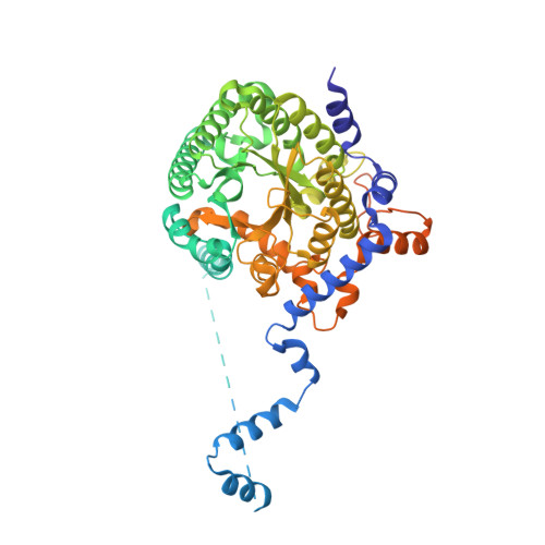
    Proline dehydrogenase (PRODH) catalyzes the first step of proline catabolism, the flavin-dependent oxidation of proline to Delta(1)-pyrroline-5-carboxylate. Here we present a structure-based study of the PRODH active site of the multifunctional Escherichia coli proline utilization A (PutA) protein using X-ray crystallography, enzyme kinetic measurements, and site-directed mutagenesis. Structures of the PutA PRODH domain complexed with competitive inhibitors acetate (K(i) = 30 mM), L-lactate (K(i) = 1 mM), and L-tetrahydro-2-furoic acid (L-THFA, K(i) = 0.2 mM) have been determined to high-resolution limits of 2.1-2.0 A. The discovery of acetate as a competitive inhibitor suggests that the carboxyl is the minimum functional group recognized by the active site, and the structures show how the enzyme exploits hydrogen-bonding and nonpolar interactions to optimize affinity for the substrate. The PRODH/L-THFA complex is the first structure of PRODH with a five-membered ring proline analogue bound in the active site and thus provides new insights into substrate recognition and the catalytic mechanism. The ring of L-THFA is nearly parallel to the middle ring of the FAD isoalloxazine, with the inhibitor C5 atom 3.3 A from the FAD N5. This geometry suggests direct hydride transfer as a plausible mechanism. Mutation of conserved active site residue Leu432 to Pro caused a 5-fold decrease in k(cat) and a severe loss in thermostability. These changes are consistent with the location of Leu432 in the hydrophobic core near residues that directly contact FAD. Our results suggest that the molecular basis for increased plasma proline levels in schizophrenic subjects carrying the missense mutation L441P is due to decreased stability of human PRODH2.


  • Organizational Affiliation
    • Department of Chemistry, University of Missouri-Columbia, Columbia, Missouri 65211, USA.

Macromolecule Content 

  • Total Structure Weight: 67.57 kDa 
  • Atom Count: 3,801 
  • Modeled Residue Count: 459 
  • Deposited Residue Count: 602 
  • Unique protein chains: 1

Macromolecules

Find similar proteins by:|  3D Structure
Entity ID: 1
MoleculeChains  Sequence LengthOrganismDetailsImage
Bifunctional putA protein602Escherichia coliMutation(s): 0 
Gene Names: PUTAPOAAB1014
EC: 1.5.99.8 (PDB Primary Data), 1.2.1.88 (UniProt), 1.5.5.2 (UniProt)
UniProt
Find proteins for P09546 (Escherichia coli (strain K12))
Explore P09546 
Go to UniProtKB:  P09546
Entity Groups
Sequence Clusters30% Identity50% Identity70% Identity90% Identity95% Identity100% Identity
UniProt GroupP09546
Sequence Annotations
Expand
Reference Sequence

Small Molecules

Ligands 2 Unique
IDChains Name / Formula / InChI Key2D Diagram3D Interactions
FAD

Query on FAD


Download:Ideal Coordinates CCD File
B [auth A]FLAVIN-ADENINE DINUCLEOTIDE
C27 H33 N9 O15 P2
VWWQXMAJTJZDQX-UYBVJOGSSA-N
TFB

Query on TFB


Download:Ideal Coordinates CCD File
C [auth A]TETRAHYDROFURAN-2-CARBOXYLIC ACID
C5 H8 O3
UJJLJRQIPMGXEZ-BYPYZUCNSA-N

Experimental Data & Validation

Experimental Data

  • Method: X-RAY DIFFRACTION
  • Resolution: 2.00 Å
  • R-Value Free:  0.253 (Depositor), 0.262 (DCC) 
  • R-Value Work:  0.214 (Depositor), 0.220 (DCC) 
  • R-Value Observed: 0.216 (Depositor) 
Space Group: I 2 2 2
Unit Cell:
Length ( Å )Angle ( ˚ )
a = 72.588α = 90
b = 141.358β = 90
c = 145.897γ = 90
Software Package:
Software NamePurpose
REFMACrefinement
HKL-2000data reduction
SCALEPACKdata scaling
CNSphasing

Structure Validation

View Full Validation Report


Ligand Structure Quality Assessment 


Entry History 

Deposition Data

Revision History  (Full details and data files)

  • Version 1.0: 2004-10-26
    Type: Initial release
  • Version 1.1: 2008-04-30
    Changes: Version format compliance
  • Version 1.2: 2011-07-13
    Changes: Advisory, Version format compliance
  • Version 1.3: 2023-08-23
    Changes: Data collection, Database references, Derived calculations, Refinement description