1THG | pdb_00001thg

1.8 ANGSTROMS REFINED STRUCTURE OF THE LIPASE FROM GEOTRICHUM CANDIDUM


Experimental Data Snapshot

  • Method: X-RAY DIFFRACTION
  • Resolution: 1.80 Å
  • R-Value Work: 
    0.157 (Depositor), 0.148 (DCC) 
  • R-Value Observed: 
    0.157 (Depositor) 

wwPDB Validation 3D Report Full Report

Validation slider image for 1THG

Ligand Structure Quality Assessment 


This is version 3.1 of the entry. See complete history

Literature

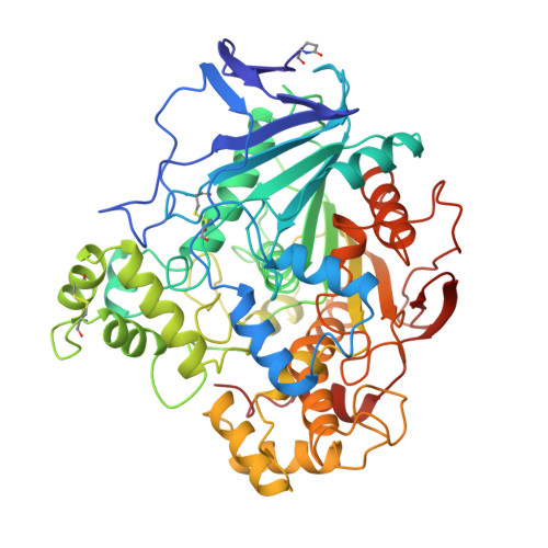
1.8 A refined structure of the lipase from Geotrichum candidum.

Schrag, J.D.Cygler, M.

(1993) J Mol Biology 230: 575-591

  • DOI: https://doi.org/10.1006/jmbi.1993.1171
  • Primary Citation Related Structures: 
    1THG

  • PubMed Abstract: 

    A lipase from the fungus Geotrichum candidum is one of only three interfacially activated lipases whose structures have been reported to date. We have previously reported the partially refined 2.2 A structure of this enzyme. We have subsequently extended the resolution and here report the fully refined 1.8 A structure of this lipase. The structure observed in the crystal is apparently not the lipolytic conformation, as the active site is not accessible from the surface of the molecule. A single large cavity is found in the interior of the molecule and extends from the catalytic Ser to two surface helices, suggesting that this face may be the region that interacts with the lipid interface. The mobility of local segments on this face is indicated by temperature factors larger than elsewhere in the molecule and by the observation of several residues whose side-chains are discretely disordered. These observations strongly suggest that this portion of the molecule is involved in interfacial and substrate binding, but the exact nature of the conformational changes induced by binding to the lipid interface can not be determined.


  • Organizational Affiliation
    • Biotechnology Research Institute, National Research Council of Canada, Montréal, Québec.

Macromolecule Content 

  • Total Structure Weight: 60.59 kDa 
  • Atom Count: 4,694 
  • Modeled Residue Count: 544 
  • Deposited Residue Count: 544 
  • Unique protein chains: 1

Macromolecules

Find similar proteins by:|  3D Structure
Entity ID: 1
MoleculeChains  Sequence LengthOrganismDetailsImage
Lipase 2544Geotrichum candidumMutation(s): 0 
Gene Names: LIP2
EC: 3.1.1.3
Membrane Entity: Yes 
UniProt
Find proteins for P22394 (Geotrichum candidum)
Explore P22394 
Go to UniProtKB:  P22394
Entity Groups
Sequence Clusters30% Identity50% Identity70% Identity90% Identity95% Identity100% Identity
UniProt GroupP22394
Glycosylation
Glycosylation Sites: 3
Sequence Annotations
Expand
Reference Sequence

Oligosaccharides

Help  
Entity ID: 2
MoleculeChains Length2D Diagram GlycosylationD Interactions
2-acetamido-2-deoxy-beta-D-glucopyranose-(1-4)-2-acetamido-2-deoxy-beta-D-glucopyranose
B
2N-Glycosylation
Glycosylation Resources
GlyTouCan: G42666HT
GlyCosmos: G42666HT
GlyGen: G42666HT

Small Molecules

Ligands 2 Unique
IDChains Name / Formula / InChI Key2D Diagram3D Interactions
NDG

Query on NDG


Download:Ideal Coordinates CCD File
D [auth A]2-acetamido-2-deoxy-alpha-D-glucopyranose
C8 H15 N O6
OVRNDRQMDRJTHS-PVFLNQBWSA-N
NAG

Query on NAG


Download:Ideal Coordinates CCD File
C [auth A]2-acetamido-2-deoxy-beta-D-glucopyranose
C8 H15 N O6
OVRNDRQMDRJTHS-FMDGEEDCSA-N
Modified Residues  1 Unique
IDChains TypeFormula2D DiagramParent
PCA
Query on PCA
A
L-PEPTIDE LINKINGC5 H7 N O3GLN

Experimental Data & Validation

Experimental Data

  • Method: X-RAY DIFFRACTION
  • Resolution: 1.80 Å
  • R-Value Work:  0.157 (Depositor), 0.148 (DCC) 
  • R-Value Observed: 0.157 (Depositor) 
Space Group: P 1 21 1
Unit Cell:
Length ( Å )Angle ( ˚ )
a = 59.4α = 90
b = 84β = 100.1
c = 56γ = 90
Software Package:
Software NamePurpose
X-PLORmodel building
X-PLORrefinement
X-PLORphasing

Structure Validation

View Full Validation Report


Ligand Structure Quality Assessment 


Entry History 

Deposition Data

Revision History  (Full details and data files)

  • Version 1.0: 1993-10-31
    Type: Initial release
  • Version 1.1: 2008-03-03
    Changes: Version format compliance
  • Version 1.2: 2011-07-13
    Changes: Non-polymer description, Version format compliance
  • Version 1.3: 2016-01-13
    Changes: Source and taxonomy
  • Version 1.4: 2017-11-29
    Changes: Derived calculations, Other
  • Version 2.0: 2019-12-25
    Changes: Database references, Derived calculations, Polymer sequence
  • Version 3.0: 2020-07-29
    Type: Remediation
    Reason: Carbohydrate remediation
    Changes: Atomic model, Data collection, Derived calculations, Structure summary
  • Version 3.1: 2024-10-23
    Changes: Data collection, Database references, Structure summary