1TFO | pdb_00001tfo

Ribonuclease from Escherichia coli complexed with its inhibitor protein


Experimental Data Snapshot

  • Method: X-RAY DIFFRACTION
  • Resolution: 2.30 Å
  • R-Value Free: 
    0.309 (Depositor) 
  • R-Value Work: 
    0.258 (Depositor) 
  • R-Value Observed: 
    0.269 (Depositor) 

wwPDB Validation 3D Report Full Report

Validation slider image for 1TFO

This is version 1.3 of the entry. See complete history

Literature

Relation between tRNase activity and the structure of colicin D according to X-ray crystallography

Yajima, S.Nakanishi, K.Takahashi, K.Ogawa, T.Hidaka, M.Kezuka, Y.Nonaka, T.Ohsawa, K.Masaki, H.

(2004) Biochem Biophys Res Commun 322: 966-973

  • DOI: https://doi.org/10.1016/j.bbrc.2004.07.206
  • Primary Citation Related Structures: 
    1TFK, 1TFO

  • PubMed Abstract: 

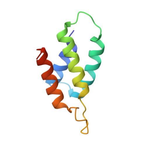
    Colicin D is a plasmid-encoded proteinaceous toxin which kills sensitive Escherichia coli. Toxicity stems from ribonuclease activity that targets exclusively four isoacceptors of tRNA(Arg) with a cleavage position between 38 and 39 of the corresponding anticodons. Since no other tRNAs with the same sequences at 38 and 39 as tRNA(Arg)s are cleaved, colicin D should be capable of recognizing some higher order structure of tRNAs. We report here two crystal structures of catalytic domains of colicin D which have different N-terminal lengths, both complexed with its cognate inhibitor protein, ImmD. A row of positive charge patches is found on the surface of the catalytic domain, suggestive of the binding site of the tRNAs. This finding, together with our refined tRNase activity experiments, indicates that the catalytic domain starting at position 595 has activity almost equivalent to that of colicin D.


  • Organizational Affiliation
    • Department of Bioscience, Tokyo University of Agriculture, Setagaya-ku, Tokyo 156-8502, Japan. yshun@nodai.ac.jp

Macromolecule Content 

  • Total Structure Weight: 22.51 kDa 
  • Atom Count: 1,561 
  • Modeled Residue Count: 189 
  • Deposited Residue Count: 194 
  • Unique protein chains: 2

Macromolecules

Find similar proteins by:|  3D Structure
Entity ID: 1
MoleculeChains  Sequence LengthOrganismDetailsImage
Colicin D103Escherichia coliMutation(s): 0 
Gene Names: CDA
UniProt
Find proteins for P17998 (Escherichia coli)
Explore P17998 
Go to UniProtKB:  P17998
Entity Groups
Sequence Clusters30% Identity50% Identity70% Identity90% Identity95% Identity100% Identity
UniProt GroupP17998
Sequence Annotations
Expand
Reference Sequence
Find similar proteins by:|  3D Structure
Entity ID: 2
MoleculeChains  Sequence LengthOrganismDetailsImage
Colicin D immunity protein91Escherichia coliMutation(s): 0 
Gene Names: CDI
UniProt
Find proteins for P11899 (Escherichia coli)
Explore P11899 
Go to UniProtKB:  P11899
Entity Groups
Sequence Clusters30% Identity50% Identity70% Identity90% Identity95% Identity100% Identity
UniProt GroupP11899
Sequence Annotations
Expand
Reference Sequence

Experimental Data & Validation

Experimental Data

  • Method: X-RAY DIFFRACTION
  • Resolution: 2.30 Å
  • R-Value Free:  0.309 (Depositor) 
  • R-Value Work:  0.258 (Depositor) 
  • R-Value Observed: 0.269 (Depositor) 
Space Group: P 41 21 2
Unit Cell:
Length ( Å )Angle ( ˚ )
a = 56.171α = 90
b = 56.171β = 90
c = 149.307γ = 90
Software Package:
Software NamePurpose
DENZOdata reduction
SCALEPACKdata scaling
CNSrefinement
CNSphasing

Structure Validation

View Full Validation Report


Entry History 

Deposition Data

Revision History  (Full details and data files)

  • Version 1.0: 2005-03-01
    Type: Initial release
  • Version 1.1: 2008-04-30
    Changes: Version format compliance
  • Version 1.2: 2011-07-13
    Changes: Version format compliance
  • Version 1.3: 2024-02-14
    Changes: Data collection, Database references