1TET | pdb_00001tet

CRYSTAL STRUCTURE OF AN ANTICHOLERA TOXIN PEPTIDE COMPLEX AT 2.3 ANGSTROMS


Experimental Data Snapshot

  • Method: X-RAY DIFFRACTION
  • Resolution: 2.30 Å
  • R-Value Work: 
    0.148 (Depositor) 
  • R-Value Observed: 
    0.148 (Depositor) 

wwPDB Validation 3D Report Full Report

Validation slider image for 1TET

This is version 1.4 of the entry. See complete history

Literature

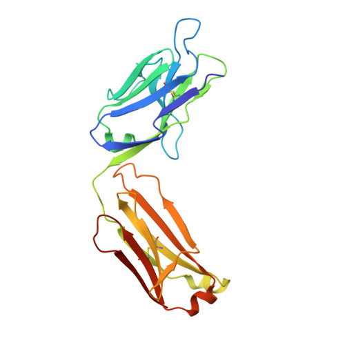
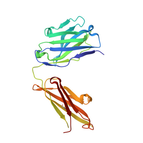
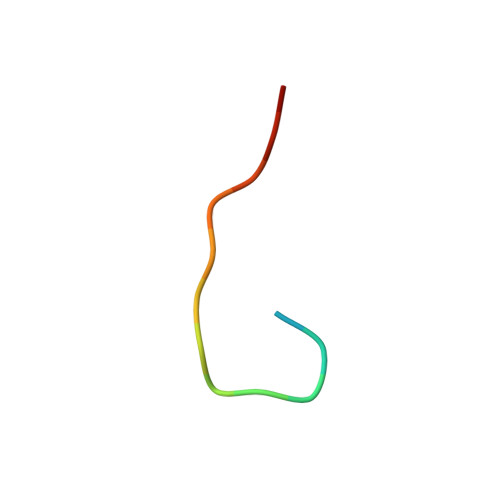
Crystal structure of an anticholera toxin peptide complex at 2.3 A.

Shoham, M.

(1993) J Mol Biology 232: 1169-1175

  • DOI: https://doi.org/10.1006/jmbi.1993.1469
  • Primary Citation Related Structures: 
    1TET

  • PubMed Abstract: 

    Cholera toxin peptide 3 (CTP3) is a 15-residue peptide corresponding in sequence to an immunogenic loop on the surface of the B-subunits of both cholera toxin and the heat-labile toxin from Escherichia coli. TE33 is the Fab fragment of a monoclonal antibody elicited against CTP3. The crystal structure of the TE33-CTP3 complex at 2.3 A resolution reveals an antigen-binding pocket, 13 A deep and 13 A wide, which is lined with many aromatic residues. The N-terminal portion of the peptide antigen CTP3 forms a type II beta-turn that fits snugly into this pocket. At gln7 the peptide backbone of CTP3 forms a kink followed by an extended C-terminal chain that seals off the cleft and buries the beta-turn underneath it. All six complementarity-determining regions of TE33 contribute to the binding of CTP3. The antibody-peptide contacts include, in addition to van der Waals' interactions and hydrogen bonds, also one salt bridge and one water molecule, which mediates the interaction.


  • Organizational Affiliation
    • Case Western Reserve University, School of Medicine, Department of Biochemistry, Cleveland, OH 44106-4935.

Macromolecule Content 

  • Total Structure Weight: 48.5 kDa 
  • Atom Count: 3,541 
  • Modeled Residue Count: 438 
  • Deposited Residue Count: 441 
  • Unique protein chains: 3

Macromolecules

Find similar proteins by:|  3D Structure
Entity ID: 1
MoleculeChains  Sequence LengthOrganismDetailsImage
IGG1 TE33 FAB (LIGHT CHAIN)A [auth L]216Mus musculusMutation(s): 0 
Find similar proteins by:|  3D Structure
Entity ID: 2
MoleculeChains  Sequence LengthOrganismDetailsImage
IGG1 TE33 FAB (HEAVY CHAIN)B [auth H]210Mus musculusMutation(s): 0 
Find similar proteins by:  Sequence   |   3D Structure  
Entity ID: 3
MoleculeChains  Sequence LengthOrganismDetailsImage
CHOLERA TOXIN PEPTIDE 3 (CTP3)C [auth P]15Escherichia coliMutation(s): 0 
Gene Names: eltB
UniProt
Find proteins for P32890 (Escherichia coli)
Explore P32890 
Go to UniProtKB:  P32890
Entity Groups
Sequence Clusters30% Identity50% Identity70% Identity90% Identity95% Identity100% Identity
UniProt GroupP32890
Sequence Annotations
Expand
Reference Sequence

Small Molecules

Ligands 1 Unique
IDChains Name / Formula / InChI Key2D Diagram3D Interactions
CIT

Query on CIT


Download:Ideal Coordinates CCD File
D [auth L]CITRIC ACID
C6 H8 O7
KRKNYBCHXYNGOX-UHFFFAOYSA-N

Experimental Data & Validation

Experimental Data

  • Method: X-RAY DIFFRACTION
  • Resolution: 2.30 Å
  • R-Value Work:  0.148 (Depositor) 
  • R-Value Observed: 0.148 (Depositor) 
Space Group: P 21 21 2
Unit Cell:
Length ( Å )Angle ( ˚ )
a = 104.15α = 90
b = 110.61β = 90
c = 40.68γ = 90
Software Package:
Software NamePurpose
X-PLORmodel building
X-PLORrefinement
X-PLORphasing

Structure Validation

View Full Validation Report


Entry History 

Deposition Data

  • Released Date: 1994-01-31 
  • Deposition Author(s): Shoham, M.

Revision History  (Full details and data files)

  • Version 1.0: 1994-01-31
    Type: Initial release
  • Version 1.1: 2008-03-03
    Changes: Version format compliance
  • Version 1.2: 2011-07-13
    Changes: Version format compliance
  • Version 1.3: 2024-06-05
    Changes: Data collection, Database references, Derived calculations, Other
  • Version 1.4: 2024-11-20
    Changes: Structure summary