1TCR | pdb_00001tcr

MURINE T-CELL ANTIGEN RECEPTOR 2C CLONE


Experimental Data Snapshot

  • Method: X-RAY DIFFRACTION
  • Resolution: 2.50 Å
  • R-Value Free: 
    0.285 (Depositor) 
  • R-Value Work: 
    0.208 (Depositor) 
  • R-Value Observed: 
    0.208 (Depositor) 

Starting Model: other
View more details

wwPDB Validation 3D Report Full Report

Validation slider image for 1TCR

This is version 2.2 of the entry. See complete history

Literature

An alphabeta T cell receptor structure at 2.5 A and its orientation in the TCR-MHC complex.

Garcia, K.C.Degano, M.Stanfield, R.L.Brunmark, A.Jackson, M.R.Peterson, P.A.Teyton, L.Wilson, I.A.

(1996) Science 274: 209-219

  • DOI: https://doi.org/10.1126/science.274.5285.209
  • Primary Citation Related Structures: 
    1TCR

  • PubMed Abstract: 

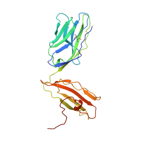
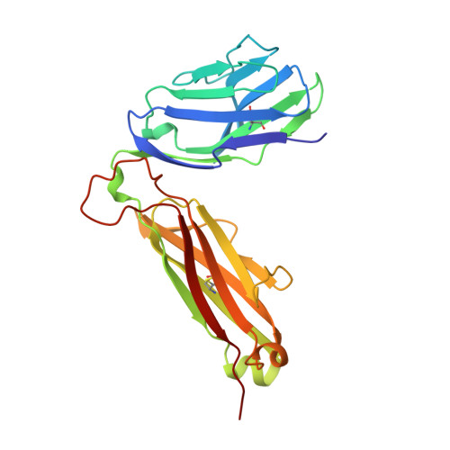
    The central event in the cellular immune response to invading microorganisms is the specific recognition of foreign peptides bound to major histocompatibility complex (MHC) molecules by the alphabeta T cell receptor (TCR). The x-ray structure of the complete extracellular fragment of a glycosylated alphabeta TCR was determined at 2.5 angstroms, and its orientation bound to a class I MHC-peptide (pMHC) complex was elucidated from crystals of the TCR-pMHC complex. The TCR resembles an antibody in the variable Valpha and Vbeta domains but deviates in the constant Calpha domain and in the interdomain pairing of Calpha with Cbeta. Four of seven possible asparagine-linked glycosylation sites have ordered carbohydrate moieties, one of which lies in the Calpha-Cbeta interface. The TCR combining site is relatively flat except for a deep hydrophobic cavity between the hypervariable CDR3s (complementarity-determining regions) of the alpha and beta chains. The 2C TCR covers the class I MHC H-2Kb binding groove so that the Valpha CDRs 1 and 2 are positioned over the amino-terminal region of the bound dEV8 peptide, the Vbeta chain CDRs 1 and 2 are over the carboxyl-terminal region of the peptide, and the Valpha and Vbeta CDR3s straddle the peptide between the helices around the central position of the peptide.


  • Organizational Affiliation
    • Department of Molecular Biology and the Skaggs Institute of Chemical Biology, Scripps Research Institute, 10550 N. Torrey Pines Road, La Jolla, CA 92037, USA.

Macromolecule Content 

  • Total Structure Weight: 50.82 kDa 
  • Atom Count: 3,589 
  • Modeled Residue Count: 439 
  • Deposited Residue Count: 439 
  • Unique protein chains: 2

Macromolecules

Find similar proteins by:|  3D Structure
Entity ID: 1
MoleculeChains  Sequence LengthOrganismDetailsImage
ALPHA, BETA T-CELL RECEPTOR (VB8.2DB2JB2.4CB2\;VA3JA58CA)202Mus musculusMutation(s): 0 
UniProt
Find proteins for P01738 (Mus musculus)
Explore P01738 
Go to UniProtKB:  P01738
Entity Groups
Sequence Clusters30% Identity50% Identity70% Identity90% Identity95% Identity100% Identity
UniProt GroupP01738
Glycosylation
Glycosylation Sites: 3Go to GlyGen: P01738-1
Sequence Annotations
Expand
Reference Sequence
Find similar proteins by:|  3D Structure
Entity ID: 2
MoleculeChains  Sequence LengthOrganismDetailsImage
ALPHA, BETA T-CELL RECEPTOR (VB8.2DB2JB2.4CB2\;VA3JA58CA)237Mus musculusMutation(s): 0 

Oligosaccharides

Help  
Entity ID: 3
MoleculeChains Length2D Diagram GlycosylationD Interactions
beta-D-mannopyranose-(1-4)-2-acetamido-2-deoxy-beta-D-glucopyranose-(1-4)-[alpha-L-fucopyranose-(1-6)]2-acetamido-2-deoxy-beta-D-glucopyranose
C
4N-Glycosylation
Glycosylation Resources
GlyTouCan: G32152BH
GlyCosmos: G32152BH
GlyGen: G32152BH
Entity ID: 4
MoleculeChains Length2D Diagram GlycosylationD Interactions
2-acetamido-2-deoxy-beta-D-glucopyranose-(1-4)-[alpha-L-fucopyranose-(1-6)]2-acetamido-2-deoxy-beta-D-glucopyranose
D
3N-Glycosylation
Glycosylation Resources
GlyTouCan: G21290RB
GlyCosmos: G21290RB
GlyGen: G21290RB
Entity ID: 5
MoleculeChains Length2D Diagram GlycosylationD Interactions
2-acetamido-2-deoxy-beta-D-glucopyranose-(1-4)-2-acetamido-2-deoxy-beta-D-glucopyranose
E
2N-Glycosylation
Glycosylation Resources
GlyTouCan: G42666HT
GlyCosmos: G42666HT
GlyGen: G42666HT

Experimental Data & Validation

Experimental Data

  • Method: X-RAY DIFFRACTION
  • Resolution: 2.50 Å
  • R-Value Free:  0.285 (Depositor) 
  • R-Value Work:  0.208 (Depositor) 
  • R-Value Observed: 0.208 (Depositor) 
Space Group: C 1 2 1
Unit Cell:
Length ( Å )Angle ( ˚ )
a = 138α = 90
b = 72.9β = 101.2
c = 57.3γ = 90
Software Package:
Software NamePurpose
DENZOdata reduction
SCALEPACKdata scaling
PHASESphasing
X-PLORmodel building
X-PLORrefinement
X-PLORphasing

Structure Validation

View Full Validation Report


Entry History 

Deposition Data

Revision History  (Full details and data files)

  • Version 1.0: 1997-03-12
    Type: Initial release
  • Version 1.1: 2008-03-24
    Changes: Version format compliance
  • Version 1.2: 2011-07-13
    Changes: Non-polymer description, Version format compliance
  • Version 1.3: 2018-03-07
    Changes: Data collection
  • Version 2.0: 2020-07-29
    Type: Remediation
    Reason: Carbohydrate remediation
    Changes: Atomic model, Data collection, Database references, Derived calculations, Structure summary
  • Version 2.1: 2024-04-03
    Changes: Data collection, Database references, Refinement description, Structure summary
  • Version 2.2: 2024-10-23
    Changes: Structure summary