1TAH | pdb_00001tah

THE CRYSTAL STRUCTURE OF TRIACYLGLYCEROL LIPASE FROM PSEUDOMONAS GLUMAE REVEALS A PARTIALLY REDUNDANT CATALYTIC ASPARTATE


Experimental Data Snapshot

  • Method: X-RAY DIFFRACTION
  • Resolution: 3.00 Å
  • R-Value Free: 
    0.226 (Depositor) 
  • R-Value Work: 
    0.159 (Depositor) 
  • R-Value Observed: 
    0.159 (Depositor) 

wwPDB Validation 3D Report Full Report

Validation slider image for 1TAH

This is version 1.4 of the entry. See complete history

Literature

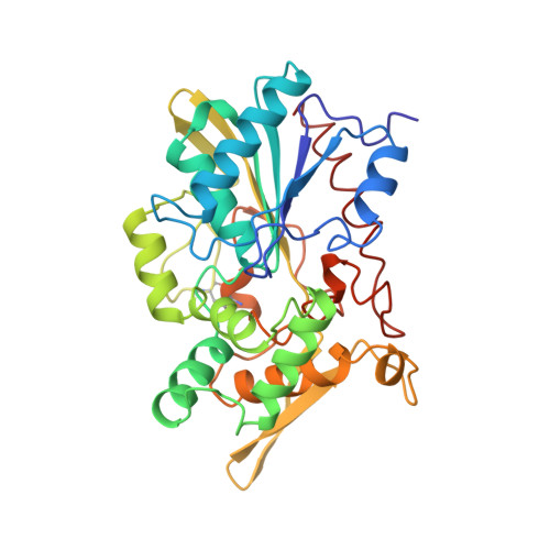
The crystal structure of triacylglycerol lipase from Pseudomonas glumae reveals a partially redundant catalytic aspartate.

Noble, M.E.Cleasby, A.Johnson, L.N.Egmond, M.R.Frenken, L.G.

(1993) FEBS Lett 331: 123-128

  • DOI: https://doi.org/10.1016/0014-5793(93)80310-q
  • Primary Citation Related Structures: 
    1TAH

  • PubMed Abstract: 

    The family of lipases (triacylglycerol-acyl-hydrolases EC 3.1.1.3) constitutes an interesting class of enzymes because of their ability to interact with lipid-water interfaces, their wide range of substrate specificities, and their potential industrial applications. Here we report the first crystal structure of a bacterial lipase, from Pseudomonas glumae. The structure is formed from three domains, the largest of which contains a subset of the alpha/beta hydrolase fold and a calcium site. Asp263, the acidic residue in the catalytic triad, has previously been mutated into an alanine with only a modest reduction in activity.


  • Organizational Affiliation
    • Laboratory of Molecular Biophysics, Department of Biochemistry, Oxford, UK.

Macromolecule Content 

  • Total Structure Weight: 132.35 kDa 
  • Atom Count: 9,320 
  • Modeled Residue Count: 1,272 
  • Deposited Residue Count: 1,272 
  • Unique protein chains: 1

Macromolecules

Find similar proteins by:|  3D Structure
Entity ID: 1
MoleculeChains  Sequence LengthOrganismDetailsImage
LIPASEA [auth B],
B [auth A],
C,
D
318Burkholderia glumaeMutation(s): 0 
EC: 3.1.1.3
UniProt
Find proteins for P0DUB8 (Burkholderia plantarii)
Explore P0DUB8 
Go to UniProtKB:  P0DUB8
Entity Groups
Sequence Clusters30% Identity50% Identity70% Identity90% Identity95% Identity100% Identity
UniProt GroupP0DUB8
Sequence Annotations
Expand
Reference Sequence

Experimental Data & Validation

Experimental Data

  • Method: X-RAY DIFFRACTION
  • Resolution: 3.00 Å
  • R-Value Free:  0.226 (Depositor) 
  • R-Value Work:  0.159 (Depositor) 
  • R-Value Observed: 0.159 (Depositor) 
Space Group: P 21 21 21
Unit Cell:
Length ( Å )Angle ( ˚ )
a = 158.16α = 90
b = 158.64β = 90
c = 63.36γ = 90
Software Package:
Software NamePurpose
X-PLORmodel building
X-PLORrefinement
X-PLORphasing

Structure Validation

View Full Validation Report


Entry History 

Deposition Data

Revision History  (Full details and data files)

  • Version 1.0: 1994-05-31
    Type: Initial release
  • Version 1.1: 2008-03-03
    Changes: Version format compliance
  • Version 1.2: 2011-07-13
    Changes: Version format compliance
  • Version 1.3: 2017-11-29
    Changes: Derived calculations, Other
  • Version 1.4: 2024-11-20
    Changes: Data collection, Database references, Derived calculations, Structure summary