1T8K | pdb_00001t8k

Crystal Structure of apo acyl carrier protein from E. coli


Experimental Data Snapshot

  • Method: X-RAY DIFFRACTION
  • Resolution: 1.10 Å
  • R-Value Free: 
    0.150 (Depositor) 
  • R-Value Work: 
    0.133 (Depositor) 
  • R-Value Observed: 
    0.134 (Depositor) 

wwPDB Validation 3D Report Full Report

Validation slider image for 1T8K

This is version 1.3 of the entry. See complete history

Literature

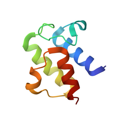
Structure of apo acyl carrier protein and a proposal to engineer protein crystallization through metal ions.

Qiu, X.Janson, C.A.

(2004) Acta Crystallogr D Biol Crystallogr 60: 1545-1554

  • DOI: https://doi.org/10.1107/S0907444904015422
  • Primary Citation Related Structures: 
    1T8K

  • PubMed Abstract: 

    A topic of current interest is engineering surface mutations in order to improve the success rate of protein crystallization. This report explores the possibility of using metal-ion-mediated crystal-packing interactions to facilitate rational design. Escherichia coli apo acyl carrier protein was chosen as a test case because of its high content of negatively charged carboxylates suitable for metal binding with moderate affinity. The protein was successfully crystallized in the presence of zinc ions. The crystal structure was determined to 1.1 A resolution with MAD phasing using anomalous signals from the co-crystallized Zn(2+) ions. The case study suggested an integrated strategy for crystallization and structure solution of proteins via engineering surface Asp and Glu mutants, crystallizing them in the presence of metal ions such as Zn(2+) and solving the structures using anomalous signals.


  • Organizational Affiliation
    • GlaxoSmithKline, King of Prussia, Pennsylvania 19406, USA. xiayang_qiu@groton.pfizer.com

Macromolecule Content 

  • Total Structure Weight: 9.58 kDa 
  • Atom Count: 750 
  • Modeled Residue Count: 77 
  • Deposited Residue Count: 77 
  • Unique protein chains: 1

Macromolecules

Find similar proteins by:|  3D Structure
Entity ID: 1
MoleculeChains  Sequence LengthOrganismDetailsImage
Acyl carrier protein77Escherichia coliMutation(s): 0 
UniProt
Find proteins for P0A6A8 (Escherichia coli (strain K12))
Explore P0A6A8 
Go to UniProtKB:  P0A6A8
Entity Groups
Sequence Clusters30% Identity50% Identity70% Identity90% Identity95% Identity100% Identity
UniProt GroupP0A6A8
Sequence Annotations
Expand
Reference Sequence

Small Molecules

Ligands 2 Unique
IDChains Name / Formula / InChI Key2D Diagram3D Interactions
IMD

Query on IMD


Download:Ideal Coordinates CCD File
L [auth A]
M [auth A]
N [auth A]
O [auth A]
P [auth A]
L [auth A],
M [auth A],
N [auth A],
O [auth A],
P [auth A],
Q [auth A]
IMIDAZOLE
C3 H5 N2
RAXXELZNTBOGNW-UHFFFAOYSA-O
ZN

Query on ZN


Download:Ideal Coordinates CCD File
B [auth A]
C [auth A]
D [auth A]
E [auth A]
F [auth A]
B [auth A],
C [auth A],
D [auth A],
E [auth A],
F [auth A],
G [auth A],
H [auth A],
I [auth A],
J [auth A],
K [auth A]
ZINC ION
Zn
PTFCDOFLOPIGGS-UHFFFAOYSA-N

Experimental Data & Validation

Experimental Data

  • Method: X-RAY DIFFRACTION
  • Resolution: 1.10 Å
  • R-Value Free:  0.150 (Depositor) 
  • R-Value Work:  0.133 (Depositor) 
  • R-Value Observed: 0.134 (Depositor) 
Space Group: P 21 21 21
Unit Cell:
Length ( Å )Angle ( ˚ )
a = 27.51α = 90
b = 41.26β = 90
c = 62.48γ = 90
Software Package:
Software NamePurpose
REFMACrefinement
DENZOdata reduction
SCALEPACKdata scaling
SOLVEphasing

Structure Validation

View Full Validation Report


Entry History 

Deposition Data

Revision History  (Full details and data files)

  • Version 1.0: 2004-09-07
    Type: Initial release
  • Version 1.1: 2008-04-30
    Changes: Version format compliance
  • Version 1.2: 2011-07-13
    Changes: Version format compliance
  • Version 1.3: 2024-02-14
    Changes: Data collection, Database references, Derived calculations