1T66

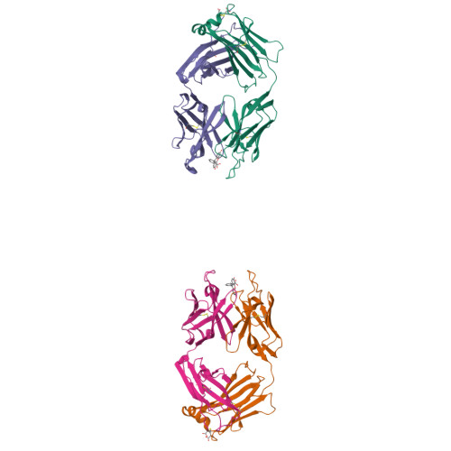
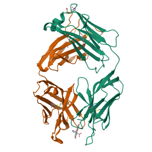
The structure of FAB with intermediate affinity for fluorescein.


Experimental Data Snapshot

  • Method: X-RAY DIFFRACTION
  • Resolution: 2.30 Å
  • R-Value Free: 0.253 
  • R-Value Work: 0.204 
  • R-Value Observed: 0.204 

Starting Model: experimental
View more details

wwPDB Validation   3D Report Full Report


Ligand Structure Quality Assessment 


This is version 1.4 of the entry. See complete history


Literature

Three-dimensional Structures of Idiotypically Related Fabs with Intermediate and High Affinity for Fluorescein.

Terzyan, S.Ramsland, P.A.Voss Jr., E.W.Herron, J.N.Edmundson, A.B.

(2004) J Mol Biol 339: 1141-1151

  • DOI: https://doi.org/10.1016/j.jmb.2004.03.080
  • Primary Citation of Related Structures:  
    1T66

  • PubMed Abstract: 

    Multi-disciplinary studies of fluorescein-protein conjugates have led to the generation of a family of antibodies with common idiotypes and affinities for fluorescein ranging over five orders of magnitude. The high affinity 4-4-20 prototype traps the ligand in a highly complementary binding slot, which is lined by multiple aromatic side-chains. An antibody (9-40) of intermediate affinity belongs to the same idiotypic family as 4-4-20 and shares substantial amino acid identities within the VL and VH domains. To establish the structural basis for the affinity differences, we solved the crystal structure of the 9-40 Fab-fluorescein complex at a resolution of 2.3A. Similar to 4-4-20, 9-40 binds fluorescein in a tight aromatic slot with its xanthenonyl ring system accommodated by end-on insertion. However, the combined effects of the amino acid substitutions have resulted in reorganization of the binding site, with the HCDR3 loops showing the greatest differences in conformations. Access to the binding site of 9-40 is substantially more open, leaving the fluorescein's phenylcarboxylate moiety partially exposed to solvent. In addition to the usage of a different D (diversity) mini-gene encoding the HCDR3 loop, the decrease in fluorescein affinity in the 9-40 antibody family appears to be correlated with the substitution of histidine (9-40) for arginine (4-4-20) in position 34 of the antibody light chains.


  • Organizational Affiliation

    Crystallography Program, Oklahoma Medical Research Foundation, Oklahoma City, OK 73104, USA.


Macromolecules
Find similar proteins by:  (by identity cutoff)  |  3D Structure
Entity ID: 1
MoleculeChains Sequence LengthOrganismDetailsImage
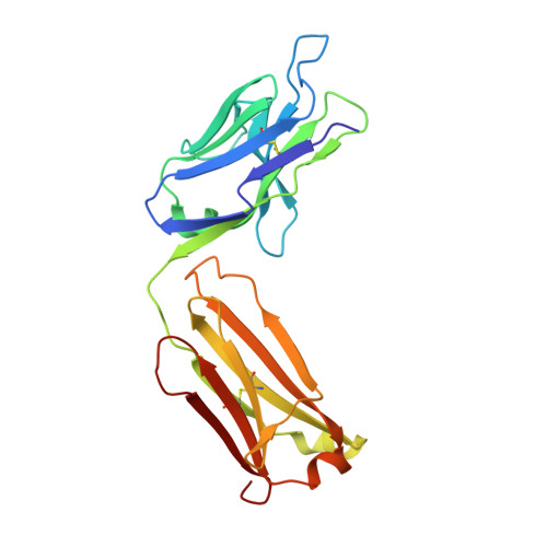
immunoglobulin light chainA [auth L],
C
219Mus musculusMutation(s): 0 
UniProt
Find proteins for Q65ZC0 (Mus musculus)
Explore Q65ZC0 
Go to UniProtKB:  Q65ZC0
Entity Groups  
Sequence Clusters30% Identity50% Identity70% Identity90% Identity95% Identity100% Identity
UniProt GroupQ65ZC0
Sequence Annotations
Expand
  • Reference Sequence
Find similar proteins by:  (by identity cutoff)  |  3D Structure
Entity ID: 2
MoleculeChains Sequence LengthOrganismDetailsImage
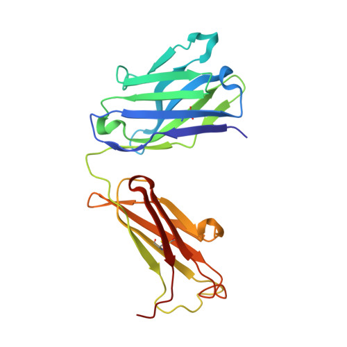
immunoglobulin heavy chainB [auth H],
D
220Mus musculusMutation(s): 0 
UniProt
Find proteins for Q91Z05 (Mus musculus)
Explore Q91Z05 
Go to UniProtKB:  Q91Z05
Entity Groups  
Sequence Clusters30% Identity50% Identity70% Identity90% Identity95% Identity100% Identity
UniProt GroupQ91Z05
Sequence Annotations
Expand
  • Reference Sequence
Small Molecules
Ligands 1 Unique
IDChains Name / Formula / InChI Key2D Diagram3D Interactions
FLU
Query on FLU

Download Ideal Coordinates CCD File 
E [auth L],
F [auth C]
2-(6-HYDROXY-3-OXO-3H-XANTHEN-9-YL)-BENZOIC ACID
C20 H12 O5
YKGGGCXBWXHKIZ-UHFFFAOYSA-N
Experimental Data & Validation

Experimental Data

  • Method: X-RAY DIFFRACTION
  • Resolution: 2.30 Å
  • R-Value Free: 0.253 
  • R-Value Work: 0.204 
  • R-Value Observed: 0.204 
  • Space Group: P 21 21 21
Unit Cell:
Length ( Å )Angle ( ˚ )
a = 61.64α = 90
b = 117.12β = 90
c = 154.48γ = 90
Software Package:
Software NamePurpose
DENZOdata reduction
SCALEPACKdata scaling
AMoREphasing
CNSrefinement

Structure Validation

View Full Validation Report


Ligand Structure Quality Assessment 


Entry History 

Deposition Data

Revision History  (Full details and data files)

  • Version 1.0: 2004-05-18
    Type: Initial release
  • Version 1.1: 2008-04-30
    Changes: Version format compliance
  • Version 1.2: 2011-07-13
    Changes: Version format compliance
  • Version 1.3: 2023-08-23
    Changes: Data collection, Database references, Derived calculations, Refinement description
  • Version 1.4: 2024-10-30
    Changes: Structure summary