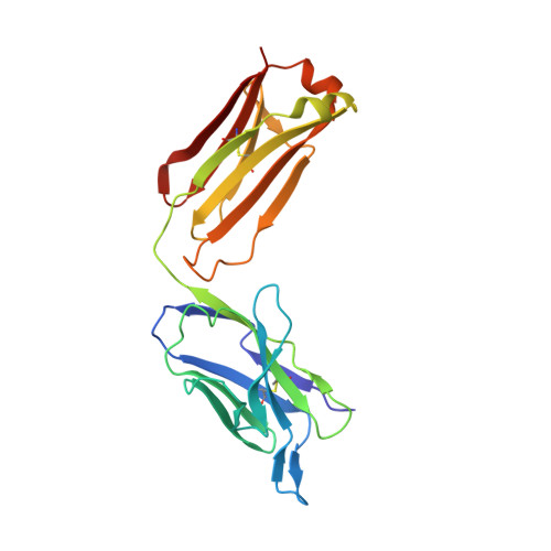
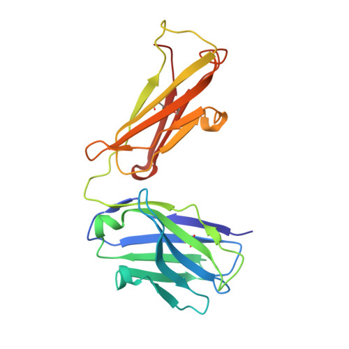
1T4K

Crystal Structure of Unliganded Aldolase Antibody 93F3 Fab


Experimental Data Snapshot

  • Method: X-RAY DIFFRACTION
  • Resolution: 2.50 Å
  • R-Value Free: 0.281 
  • R-Value Work: 0.233 
  • R-Value Observed: 0.233 

Starting Model: experimental
View more details

wwPDB Validation   3D Report Full Report


This is version 1.4 of the entry. See complete history


Literature

The Origin of Enantioselectivity in Aldolase Antibodies: Crystal Structure, Site-directed Mutagenesis, and Computational Analysis

Zhu, X.Tanaka, F.Hu, Y.Heine, A.Fuller, R.Zhong, G.Olson, A.J.Lerner, R.A.Barbas, C.F.Wilson, I.A.

(2004) J Mol Biol 343: 1269-1280

  • DOI: https://doi.org/10.1016/j.jmb.2004.08.102
  • Primary Citation of Related Structures:  
    1T4K

  • PubMed Abstract: 

    Catalytic aldolase antibodies, generated by reactive immunization, catalyze the aldol reaction with the efficiency of natural enzymes, but accept a much broader range of substrates. Two separate groups of aldolase antibodies that catalyze the same aldol reactions with antipodal selectivity were analyzed by comparing their amino acid sequences with their crystal structures, site-directed mutagenesis data, and computational docking of the transition states of the aldol reaction. The crystal structure of aldolase antibody 93F3 Fab' at 2.5A resolution revealed a combining site with two lysine residues, including LysL89 that reacts to form the covalent enamine intermediate. In contrast, antibody 33F12 has one active site lysine, LysH93. The reactive lysine residues in each group of antibodies are differentially located on the heavy and light chain variable regions in pseudo-symmetric opposite orientations, but both within highly hydrophobic environments. Thus, the defining feature for the observed enantioselectivities of these aldolase antibody catalysts is the respective location and relative disposition of the reactive lysine residues within the active sites of these catalysts.


  • Organizational Affiliation

    Department of Molecular Biology, The Scripps Research Institute, 10550 North Torrey Pines Road, La Jolla, CA 92037, USA.


Macromolecules
Find similar proteins by:  (by identity cutoff)  |  3D Structure
Entity ID: 1
MoleculeChains Sequence LengthOrganismDetailsImage
IMMUNOGLOBULIN IGG1, KAPPA LIGHT CHAIN
A, C
217Mus musculusMutation(s): 0 
Entity Groups  
Sequence Clusters30% Identity50% Identity70% Identity90% Identity95% Identity100% Identity
Sequence Annotations
Expand
  • Reference Sequence
Find similar proteins by:  (by identity cutoff)  |  3D Structure
Entity ID: 2
MoleculeChains Sequence LengthOrganismDetailsImage
IMMUNOGLOBULIN IGG1, HEAVY CHAIN
B, D
217Mus musculusMutation(s): 0 
Entity Groups  
Sequence Clusters30% Identity50% Identity70% Identity90% Identity95% Identity100% Identity
Sequence Annotations
Expand
  • Reference Sequence
Experimental Data & Validation

Experimental Data

  • Method: X-RAY DIFFRACTION
  • Resolution: 2.50 Å
  • R-Value Free: 0.281 
  • R-Value Work: 0.233 
  • R-Value Observed: 0.233 
  • Space Group: P 21 21 21
Unit Cell:
Length ( Å )Angle ( ˚ )
a = 79.701α = 90
b = 81.228β = 90
c = 149.496γ = 90
Software Package:
Software NamePurpose
DENZOdata reduction
SCALEPACKdata scaling
AMoREphasing
CNSrefinement

Structure Validation

View Full Validation Report


Entry History 

Deposition Data

Revision History  (Full details and data files)

  • Version 1.0: 2004-11-02
    Type: Initial release
  • Version 1.1: 2008-04-30
    Changes: Version format compliance
  • Version 1.2: 2011-07-13
    Changes: Version format compliance
  • Version 1.3: 2023-08-23
    Changes: Data collection, Database references, Derived calculations, Refinement description
  • Version 1.4: 2024-10-30
    Changes: Structure summary