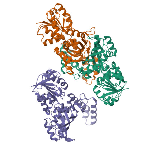
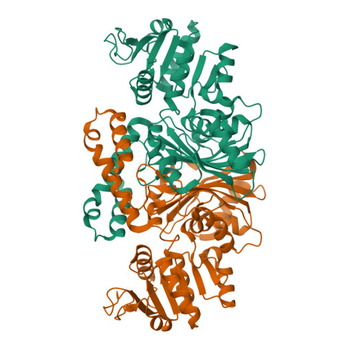
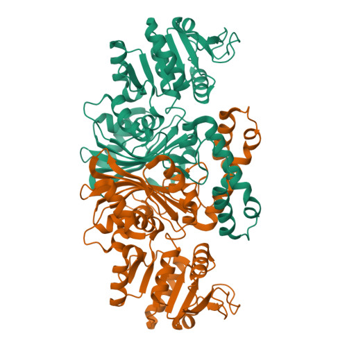
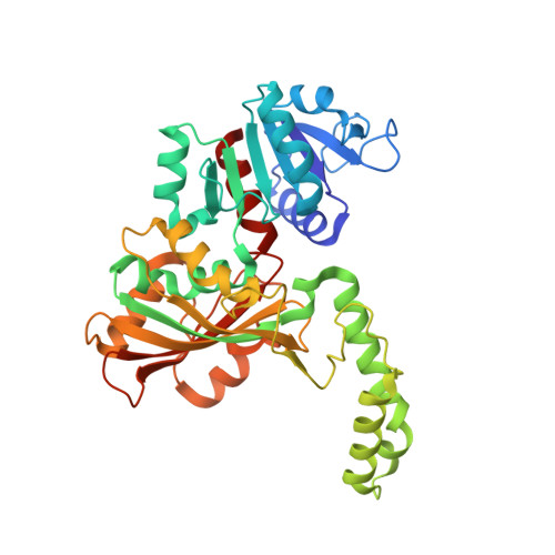
1T4D

Crystal structure of Escherichia coli aspartate beta-semialdehyde dehydrogenase (EcASADH), at 1.95 Angstrom resolution


Experimental Data Snapshot

  • Method: X-RAY DIFFRACTION
  • Resolution: 1.95 Å
  • R-Value Free: 0.250 
  • R-Value Work: 0.204 
  • R-Value Observed: 0.204 

Starting Model: experimental
View more details

wwPDB Validation   3D Report Full Report


This is version 1.5 of the entry. See complete history


Literature

High-resolution Structures Reveal Details of Domain Closure and "Half-of-sites-reactivity" in Escherichia coli Aspartate beta-Semialdehyde Dehydrogenase.

Nichols, C.E.Dhaliwal, B.Lockyer, M.Hawkins, A.R.Stammers, D.K.

(2004) J Mol Biol 341: 797-806

  • DOI: https://doi.org/10.1016/j.jmb.2004.05.073
  • Primary Citation of Related Structures:  
    1T4B, 1T4D

  • PubMed Abstract: 

    Two high-resolution structures have been determined for Eschericia coli aspartate beta-semialdehyde dehydrogenase (ecASADH), an enzyme of the aspartate biosynthetic pathway, which is a potential target for novel antimicrobial drugs. Both ASADH structures were of the open form and were refined to 1.95 A and 1.6 A resolution, allowing a more detailed comparison with the closed form of the enzyme than previously possible. A more complex scheme for domain closure is apparent with the subunit being split into two further sub-domains with relative motions about three hinge axes. Analysis of hinge data and torsion-angle difference plots is combined to allow the proposal of a detailed structural mechanism for ecASADH domain closure. Additionally, asymmetric distortions of individual subunits are identified, which form the basis for the previously reported "half-of-the-sites reactivity" (HOSR). A putative explanation of this arrangement is also presented, suggesting the HOSR system may provide a means for ecASADH to offset the energy required to remobilise flexible loops at the end of the reaction cycle, and hence avoid falling into an energy minimum.


  • Organizational Affiliation

    Division of Structural Biology, The Wellcome Trust Centre for Human Genetics, University of Oxford, Roosevelt Drive, Oxford, OX3 7BN, UK.


Macromolecules
Find similar proteins by:  (by identity cutoff)  |  3D Structure
Entity ID: 1
MoleculeChains Sequence LengthOrganismDetailsImage
Aspartate-semialdehyde dehydrogenase
A, B, C
367Escherichia coliMutation(s): 1 
Gene Names: ASDHOMB3433Z4797ECS4278SF3456S4307
EC: 1.2.1.11
UniProt
Find proteins for P0A9Q9 (Escherichia coli (strain K12))
Explore P0A9Q9 
Go to UniProtKB:  P0A9Q9
Entity Groups  
Sequence Clusters30% Identity50% Identity70% Identity90% Identity95% Identity100% Identity
UniProt GroupP0A9Q9
Sequence Annotations
Expand
  • Reference Sequence
Experimental Data & Validation

Experimental Data

  • Method: X-RAY DIFFRACTION
  • Resolution: 1.95 Å
  • R-Value Free: 0.250 
  • R-Value Work: 0.204 
  • R-Value Observed: 0.204 
  • Space Group: P 21 21 2
Unit Cell:
Length ( Å )Angle ( ˚ )
a = 172.42α = 90
b = 117.21β = 90
c = 57.7γ = 90
Software Package:
Software NamePurpose
CNSrefinement
DENZOdata reduction
SCALEPACKdata scaling
CNSphasing

Structure Validation

View Full Validation Report


Entry History 

Deposition Data

Revision History  (Full details and data files)

  • Version 1.0: 2004-08-17
    Type: Initial release
  • Version 1.1: 2008-04-30
    Changes: Version format compliance
  • Version 1.2: 2011-07-13
    Changes: Version format compliance
  • Version 1.3: 2011-11-16
    Changes: Atomic model
  • Version 1.4: 2021-10-27
    Changes: Database references
  • Version 1.5: 2023-08-23
    Changes: Data collection, Refinement description