1SQL | pdb_00001sql

Crystal structure of 7,8-dihydroneopterin aldolase in complex with guanine


Experimental Data Snapshot

  • Method: X-RAY DIFFRACTION
  • Resolution: 2.20 Å
  • R-Value Free: 
    0.260 (Depositor), 0.258 (DCC) 
  • R-Value Work: 
    0.204 (Depositor), 0.195 (DCC) 

Starting Model: experimental
View more details

wwPDB Validation 3D Report Full Report

Validation slider image for 1SQL

Ligand Structure Quality Assessment 


This is version 1.3 of the entry. See complete history

Literature

Biosynthesis of Tetrahydrofolate in Plants: Crystal Structure of 7,8-Dihydroneopterin Aldolase from Arabidopsis thaliana Reveals a Novel Adolase Class.

Bauer, S.Schott, A.K.Illarionova, V.Bacher, A.Huber, R.Fischer, M.

(2004) J Mol Biology 339: 967-979

  • DOI: https://doi.org/10.1016/j.jmb.2004.04.034
  • Primary Citation Related Structures: 
    1SQL

  • PubMed Abstract: 

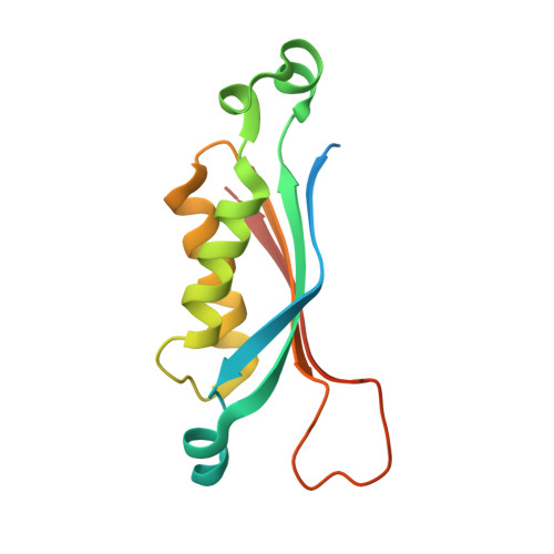
    Dihydroneopterin aldolase (DHNA) catalyses a retroaldol reaction yielding 6-hydroxymethyl-7,8-dihydropterin, a biosynthetic precursor of the vitamin, tetrahydrofolate. The enzyme is a potential target for antimicrobial and anti-parasite chemotherapy. A gene specifying a dihydroneopterin aldolase from Arabidopsis thaliana was expressed in a recombinant Escherichia coli strain. The recombinant protein was purified to apparent homogeneity and crystallised using polyethylenglycol as the precipitating agent. The crystal structure was solved by X-ray diffraction analysis at 2.2A resolution. The enzyme forms a D(4)-symmetric homooctamer. Each polypeptide chain is folded into a single domain comprising an antiparallel four-stranded beta-sheet and two long alpha-helices. Four monomers are arranged in a tetrameric ring, and two of these rings form a hollow cylinder. Well defined purine derivatives are found at all eight topologically equivalent active sites. The subunit fold of the enzyme is related to substructures of dihydroneopterin triphosphate epimerase, GTP cyclohydrolase I, and pyruvoyltetrahydropterin synthase, which are all involved in the biosynthesis of pteridine type cofactors, and to urate oxidase, although some members of that superfamily have no detectable sequence similarity. Due to structural and mechanistical differences of DHNA in comparison with class I and class II aldolases, a new aldolase class is proposed.


  • Organizational Affiliation
    • Max-Planck-Institut für Biochemie, Abteilung Strukturforschung, Am Klopferspitz 18a, D-82152 Martinsried, Germany. stbauer@biochem.mpg.de

Macromolecule Content 

  • Total Structure Weight: 263.26 kDa 
  • Atom Count: 16,400 
  • Modeled Residue Count: 1,939 
  • Deposited Residue Count: 2,336 
  • Unique protein chains: 1

Macromolecules

Find similar proteins by:|  3D Structure
Entity ID: 1
MoleculeChains  Sequence LengthOrganismDetailsImage
dihydroneopterin aldolase
A, B, C, D, E
A, B, C, D, E, F, G, H, I, J, K, L, M, N, O, P
146Arabidopsis thalianaMutation(s): 0 
EC: 4.1.2.25
UniProt
Find proteins for Q9SF23 (Arabidopsis thaliana)
Explore Q9SF23 
Go to UniProtKB:  Q9SF23
Entity Groups
Sequence Clusters30% Identity50% Identity70% Identity90% Identity95% Identity100% Identity
UniProt GroupQ9SF23
Sequence Annotations
Expand
Reference Sequence

Small Molecules

Ligands 1 Unique
IDChains Name / Formula / InChI Key2D Diagram3D Interactions
GUN

Query on GUN


Download:Ideal Coordinates CCD File
AA [auth J]
BA [auth L]
CA [auth M]
DA [auth O]
EA [auth P]
AA [auth J],
BA [auth L],
CA [auth M],
DA [auth O],
EA [auth P],
FA [auth P],
Q [auth A],
R [auth B],
S [auth B],
T [auth D],
U [auth F],
V [auth G],
W [auth G],
X [auth H],
Y [auth I],
Z [auth J]
GUANINE
C5 H5 N5 O
UYTPUPDQBNUYGX-UHFFFAOYSA-N

Experimental Data & Validation

Experimental Data

  • Method: X-RAY DIFFRACTION
  • Resolution: 2.20 Å
  • R-Value Free:  0.260 (Depositor), 0.258 (DCC) 
  • R-Value Work:  0.204 (Depositor), 0.195 (DCC) 
Space Group: P 1
Unit Cell:
Length ( Å )Angle ( ˚ )
a = 63.55α = 90.14
b = 84.22β = 89.99
c = 89.05γ = 76.17
Software Package:
Software NamePurpose
DENZOdata reduction
SCALEPACKdata scaling
AMoREphasing
CNSrefinement

Structure Validation

View Full Validation Report


Ligand Structure Quality Assessment 


Entry History 

Deposition Data

Revision History  (Full details and data files)

  • Version 1.0: 2004-06-08
    Type: Initial release
  • Version 1.1: 2008-04-29
    Changes: Version format compliance
  • Version 1.2: 2011-07-13
    Changes: Version format compliance
  • Version 1.3: 2023-08-23
    Changes: Data collection, Database references, Derived calculations, Refinement description