1SQ5 | pdb_00001sq5

Crystal Structure of E. coli Pantothenate kinase


Experimental Data Snapshot

  • Method: X-RAY DIFFRACTION
  • Resolution: 2.20 Å
  • R-Value Free: 
    0.225 (Depositor), 0.240 (DCC) 
  • R-Value Work: 
    0.187 (Depositor), 0.202 (DCC) 
  • R-Value Observed: 
    0.187 (Depositor) 

wwPDB Validation 3D Report Full Report

Validation slider image for 1SQ5

Ligand Structure Quality Assessment 


This is version 1.3 of the entry. See complete history

Literature

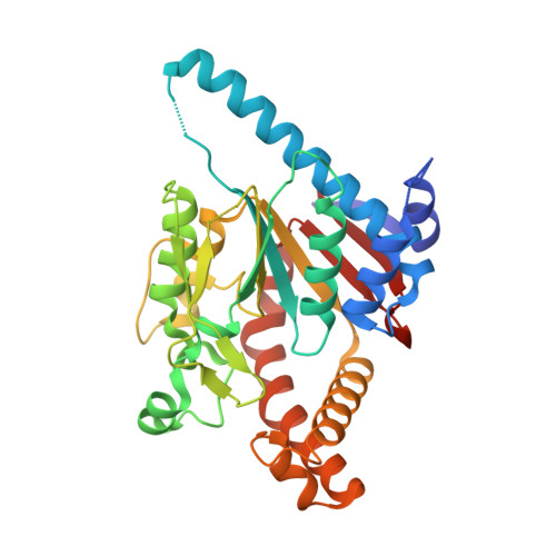
The structure of the pantothenate kinase.ADP.pantothenate ternary complex reveals the relationship between the binding sites for substrate, allosteric regulator, and antimetabolites.

Ivey, R.A.Zhang, Y.-M.Virga, K.G.Hevener, K.Lee, R.E.Rock, C.O.Jackowski, S.Park, H.-W.

(2004) J Biological Chem 279: 35622-35629

  • DOI: https://doi.org/10.1074/jbc.M403152200
  • Primary Citation Related Structures: 
    1SQ5

  • PubMed Abstract: 

    Pantothenate kinase catalyzes the first step in the biosynthesis of coenzyme A, the major acyl group carrier in biology. In bacteria, regulation of pantothenate kinase activity is a major factor in controlling intracellular coenzyme A levels, and pantothenate analogs are growth-inhibiting antimetabolites. We have extended the structural information on Escherichia coli pantothenate kinase by determining the structure of the enzyme.ADP. pantothenate ternary complex. Pantothenate binding induces a significant conformational change in amino acids 243-263, which form a "lid" that folds over the open pantothenate binding groove. The positioning of the substrates suggests the reaction proceeds by a concerted mechanism that involves a dissociative transition state, although the negative charge neutralization of the gamma-phosphate by Arg-243, Lys-101, and Mg(2+) coupled with hydrogen bonding of the C1 of pantothenate to Asp-127 suggests different interpretations of the phosphoryl transfer mechanism of pantothenate kinase. N-alkylpantothenamides are substrates for pantothenate kinase. Modeling these antimetabolites into the pantothenate active site predicts that they bind in the same orientation as pantothenate with their alkyl chains interacting with the hydrophobic dome over the pantothenate pocket, which is also accessed by the beta-mercaptoethylamine moiety of the allosteric regulator, coenzyme A. These structural/biochemical studies illustrate the intimate relationship between the substrate, allosteric regulator, and antimetabolite binding sites on pantothenate kinase and provide a framework for studies of its catalysis and feedback regulation.


  • Organizational Affiliation
    • Department of Structural Biology, St. Jude Children's Research Hospital, Memphis, Tennessee 38105, USA.

Macromolecule Content 

  • Total Structure Weight: 144.48 kDa 
  • Atom Count: 10,703 
  • Modeled Residue Count: 1,206 
  • Deposited Residue Count: 1,232 
  • Unique protein chains: 1

Macromolecules

Find similar proteins by:|  3D Structure
Entity ID: 1
MoleculeChains  Sequence LengthOrganismDetailsImage
Pantothenate kinase
A, B, C, D
308Escherichia coliMutation(s): 0 
Gene Names: COAARTSPANKB3974C4933Z5545ECS4901
EC: 2.7.1.33
UniProt
Find proteins for P0A6I3 (Escherichia coli (strain K12))
Explore P0A6I3 
Go to UniProtKB:  P0A6I3
Entity Groups
Sequence Clusters30% Identity50% Identity70% Identity90% Identity95% Identity100% Identity
UniProt GroupP0A6I3
Sequence Annotations
Expand
Reference Sequence

Small Molecules

Ligands 2 Unique
IDChains Name / Formula / InChI Key2D Diagram3D Interactions
ADP

Query on ADP


Download:Ideal Coordinates CCD File
F [auth A],
H [auth B],
J [auth C],
L [auth D]
ADENOSINE-5'-DIPHOSPHATE
C10 H15 N5 O10 P2
XTWYTFMLZFPYCI-KQYNXXCUSA-N
PAU

Query on PAU


Download:Ideal Coordinates CCD File
E [auth A],
G [auth B],
I [auth C],
K [auth D]
PANTOTHENOIC ACID
C9 H17 N O5
GHOKWGTUZJEAQD-ZETCQYMHSA-N

Experimental Data & Validation

Experimental Data

  • Method: X-RAY DIFFRACTION
  • Resolution: 2.20 Å
  • R-Value Free:  0.225 (Depositor), 0.240 (DCC) 
  • R-Value Work:  0.187 (Depositor), 0.202 (DCC) 
  • R-Value Observed: 0.187 (Depositor) 
Space Group: C 1 2 1
Unit Cell:
Length ( Å )Angle ( ˚ )
a = 181.203α = 90
b = 181.655β = 104.79
c = 47.418γ = 90
Software Package:
Software NamePurpose
CNSrefinement
DENZOdata reduction
SCALEPACKdata scaling

Structure Validation

View Full Validation Report


Ligand Structure Quality Assessment 


Entry History 

Deposition Data

Revision History  (Full details and data files)

  • Version 1.0: 2004-09-28
    Type: Initial release
  • Version 1.1: 2008-04-29
    Changes: Version format compliance
  • Version 1.2: 2011-07-13
    Changes: Version format compliance
  • Version 1.3: 2024-02-14
    Changes: Data collection, Database references, Derived calculations