1SOA | pdb_00001soa

Human DJ-1 with sulfinic acid


Experimental Data Snapshot

  • Method: X-RAY DIFFRACTION
  • Resolution: 1.20 Å
  • R-Value Free: 
    0.141 (Depositor), 0.132 (DCC) 
  • R-Value Work: 
    0.113 (Depositor), 0.108 (DCC) 
  • R-Value Observed: 
    0.113 (Depositor) 

Starting Model: experimental
View more details

wwPDB Validation 3D Report Full Report

Validation slider image for 1SOA

This is version 1.6 of the entry. See complete history

Literature

The Parkinson's disease protein DJ-1 is neuroprotective due to cysteine-sulfinic acid-driven mitochondrial localization.

Wilson, M.A.Miller, D.W.Ahmad, R.McLendon, C.Bandyopadhyay, S.Baptista, M.J.Ringe, D.Petsko, G.A.Cookson, M.R.

(2004) Proc Natl Acad Sci U S A 101: 9103-9108

  • DOI: https://doi.org/10.1073/pnas.0402959101
  • Primary Citation Related Structures: 
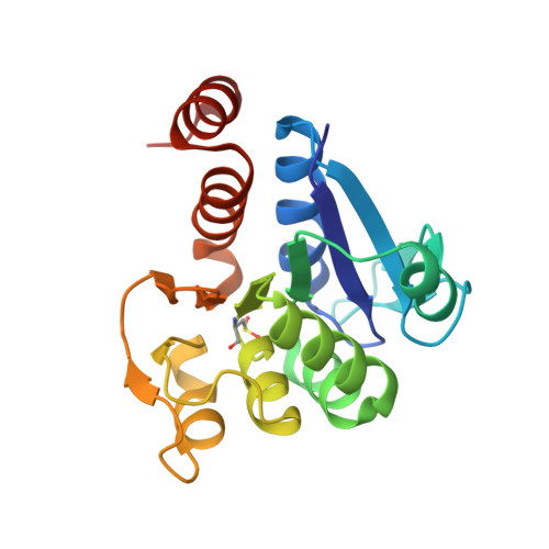
    1SOA

  • PubMed Abstract: 

    Loss-of-function DJ-1 mutations can cause early-onset Parkinson's disease. The function of DJ-1 is unknown, but an acidic isoform accumulates after oxidative stress, leading to the suggestion that DJ-1 is protective under these conditions. We addressed whether this represents a posttranslational modification at cysteine residues by systematically mutating cysteine residues in human DJ-1. WT or C53A DJ-1 was readily oxidized in cultured cells, generating a pI 5.8 isoform, but an artificial C106A mutant was not. We observed a cysteine-sulfinic acid at C106 in crystalline DJ-1 but no modification of C53 or C46. Oxidation of DJ-1 was promoted by the crystallization procedure. In addition, oxidation-induced mitochondrial relocalization of DJ-1 and protection against cell death were abrogated in C106A but not C53A or C46A. We suggest that DJ-1 protects against neuronal death, and that this is signaled by acidification of the key cysteine residue, C106.


  • Organizational Affiliation
    • Laboratory of Neurogenetics, National Institute on Aging, 9000 Rockville Pike, Bethesda, MD 20892-1589, USA.

Macromolecule Content 

  • Total Structure Weight: 19.95 kDa 
  • Atom Count: 1,778 
  • Modeled Residue Count: 187 
  • Deposited Residue Count: 189 
  • Unique protein chains: 1

Macromolecules

Find similar proteins by:|  3D Structure
Entity ID: 1
MoleculeChains  Sequence LengthOrganismDetailsImage
RNA-binding protein regulatory subunit; oncogene DJ1189Homo sapiensMutation(s): 1 
EC: 3.5.1 (UniProt), 3.1.2 (UniProt), 3.5.1.124 (UniProt)
UniProt & NIH Common Fund Data Resources
Find proteins for Q99497 (Homo sapiens)
Explore Q99497 
Go to UniProtKB:  Q99497
PHAROS:  Q99497
GTEx:  ENSG00000116288 
Entity Groups
Sequence Clusters30% Identity50% Identity70% Identity90% Identity95% Identity100% Identity
UniProt GroupQ99497
Sequence Annotations
Expand
Reference Sequence

Small Molecules

Modified Residues  1 Unique
IDChains TypeFormula2D DiagramParent
CSD
Query on CSD
A
L-PEPTIDE LINKINGC3 H7 N O4 SCYS

Experimental Data & Validation

Experimental Data

  • Method: X-RAY DIFFRACTION
  • Resolution: 1.20 Å
  • R-Value Free:  0.141 (Depositor), 0.132 (DCC) 
  • R-Value Work:  0.113 (Depositor), 0.108 (DCC) 
  • R-Value Observed: 0.113 (Depositor) 
Space Group: P 31 2 1
Unit Cell:
Length ( Å )Angle ( ˚ )
a = 75.13α = 90
b = 75.13β = 90
c = 75.094γ = 120
Software Package:
Software NamePurpose
SHELXmodel building
SHELXL-97refinement
DENZOdata reduction
SCALEPACKdata scaling
SHELXL-97phasing

Structure Validation

View Full Validation Report


Entry History 

Deposition Data

Revision History  (Full details and data files)

  • Version 1.0: 2004-06-22
    Type: Initial release
  • Version 1.1: 2008-04-29
    Changes: Version format compliance
  • Version 1.2: 2011-07-13
    Changes: Derived calculations, Version format compliance
  • Version 1.3: 2014-11-12
    Changes: Non-polymer description
  • Version 1.4: 2017-10-11
    Changes: Refinement description
  • Version 1.5: 2023-08-23
    Changes: Data collection, Database references, Derived calculations, Refinement description
  • Version 1.6: 2024-10-30
    Changes: Structure summary