1SLQ | pdb_00001slq

Crystal structure of the trimeric state of the rhesus rotavirus VP4 membrane interaction domain, VP5CT


Experimental Data Snapshot

  • Method: X-RAY DIFFRACTION
  • Resolution: 3.20 Å
  • R-Value Free: 
    0.338 (Depositor), 0.344 (DCC) 
  • R-Value Work: 
    0.308 (Depositor), 0.313 (DCC) 
  • R-Value Observed: 
    0.310 (Depositor) 

wwPDB Validation 3D Report Full Report

Validation slider image for 1SLQ

This is version 1.3 of the entry. See complete history

Literature

Structural rearrangements in the membrane penetration protein of a non-enveloped virus.

Dormitzer, P.R.Nason, E.B.Prasad, B.V.Harrison, S.C.

(2004) Nature 430: 1053-1058

  • DOI: https://doi.org/10.1038/nature02836
  • Primary Citation Related Structures: 
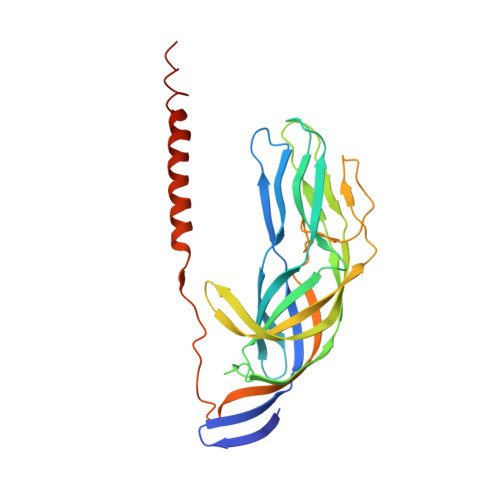
    1SLQ

  • PubMed Abstract: 

    Non-enveloped virus particles (those that lack a lipid-bilayer membrane) must breach the membrane of a target host cell to gain access to its cytoplasm. So far, the molecular mechanism of this membrane penetration step has resisted structural analysis. The spike protein VP4 is a principal component in the entry apparatus of rotavirus, a non-enveloped virus that causes gastroenteritis and kills 440,000 children each year. Trypsin cleavage of VP4 primes the virus for entry by triggering a rearrangement that rigidifies the VP4 spikes. We have determined the crystal structure, at 3.2 A resolution, of the main part of VP4 that projects from the virion. The crystal structure reveals a coiled-coil stabilized trimer. Comparison of this structure with the two-fold clustered VP4 spikes in a approximately 12 A resolution image reconstruction from electron cryomicroscopy of trypsin-primed virions shows that VP4 also undergoes a second rearrangement, in which the oligomer reorganizes and each subunit folds back on itself, translocating a potential membrane-interaction peptide from one end of the spike to the other. This rearrangement resembles the conformational transitions of membrane fusion proteins of enveloped viruses.


  • Organizational Affiliation
    • Department of Pediatrics, Harvard Medical School, and the Laboratory of Molecular Medicine, Children's Hospital, Boston, Massachusetts 02115, USA. dormitze@crystal.harvard.edu

Macromolecule Content 

  • Total Structure Weight: 187.85 kDa 
  • Atom Count: 12,772 
  • Modeled Residue Count: 1,604 
  • Deposited Residue Count: 1,668 
  • Unique protein chains: 1

Macromolecules

Find similar proteins by:|  3D Structure
Entity ID: 1
MoleculeChains  Sequence LengthOrganismDetailsImage
VP4
A, B, C, D, E
A, B, C, D, E, F
278Simian rotavirus A strain RRVMutation(s): 0 
Gene Names: gene segment 4
UniProt
Find proteins for P12473 (Rotavirus A (strain RVA/Monkey/United States/RRV/1975/G3P5B[3]))
Explore P12473 
Go to UniProtKB:  P12473
Entity Groups
Sequence Clusters30% Identity50% Identity70% Identity90% Identity95% Identity100% Identity
UniProt GroupP12473
Sequence Annotations
Expand
Reference Sequence

Experimental Data & Validation

Experimental Data

  • Method: X-RAY DIFFRACTION
  • Resolution: 3.20 Å
  • R-Value Free:  0.338 (Depositor), 0.344 (DCC) 
  • R-Value Work:  0.308 (Depositor), 0.313 (DCC) 
  • R-Value Observed: 0.310 (Depositor) 
Space Group: P 42 2 2
Unit Cell:
Length ( Å )Angle ( ˚ )
a = 244.843α = 90
b = 244.843β = 90
c = 104.778γ = 90
Software Package:
Software NamePurpose
HKL-2000data collection
SCALEPACKdata scaling
MLPHAREphasing
CNSrefinement
HKL-2000data reduction

Structure Validation

View Full Validation Report


Entry History 

Deposition Data

Revision History  (Full details and data files)

  • Version 1.0: 2004-08-31
    Type: Initial release
  • Version 1.1: 2008-04-29
    Changes: Version format compliance
  • Version 1.2: 2011-07-13
    Changes: Version format compliance
  • Version 1.3: 2024-02-14
    Changes: Data collection, Database references