1SIG | pdb_00001sig

CRYSTAL STRUCTURE OF A SIGMA70 SUBUNIT FRAGMENT FROM ESCHERICHIA COLI RNA POLYMERASE


Experimental Data Snapshot

  • Method: X-RAY DIFFRACTION
  • Resolution: 2.60 Å
  • R-Value Free: 
    0.315 (Depositor) 
  • R-Value Work: 
    0.218 (Depositor), 0.223 (DCC) 
  • R-Value Observed: 
    0.218 (Depositor) 

wwPDB Validation 3D Report Full Report

Validation slider image for 1SIG

This is version 1.3 of the entry. See complete history

Literature

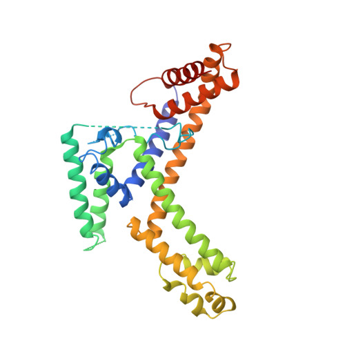
Crystal structure of a sigma 70 subunit fragment from E. coli RNA polymerase.

Malhotra, A.Severinova, E.Darst, S.A.

(1996) Cell 87: 127-136

  • DOI: https://doi.org/10.1016/s0092-8674(00)81329-x
  • Primary Citation Related Structures: 
    1SIG

  • PubMed Abstract: 

    The 2.6 A crystal structure of a fragment of the sigma 70 promoter specificity subunit of E. coli RNA polymerase is described. Residues involved in core RNA polymerase binding lie on one face of the structure. On the opposite face, aligned along one helix, are exposed residues that interact with the -10 consensus promoter element (the Pribnow box), including four aromatic residues involved in promoter melting. The structure suggests one way in which DNA interactions may be inhibited in the absence of RNA polymerase and provides a framework for the interpretation of a large number of genetic and biochemical analyses.


  • Organizational Affiliation
    • Rockefeller University, New York, New York 10021, USA.

Macromolecule Content 

  • Total Structure Weight: 39.09 kDa 
  • Atom Count: 2,593 
  • Modeled Residue Count: 305 
  • Deposited Residue Count: 339 
  • Unique protein chains: 1

Macromolecules

Find similar proteins by:|  3D Structure
Entity ID: 1
MoleculeChains  Sequence LengthOrganismDetailsImage
RNA POLYMERASE PRIMARY SIGMA FACTOR339Escherichia coli K-12Mutation(s): 0 
Gene Names: RPOD
UniProt
Find proteins for P00579 (Escherichia coli (strain K12))
Explore P00579 
Go to UniProtKB:  P00579
Entity Groups
Sequence Clusters30% Identity50% Identity70% Identity90% Identity95% Identity100% Identity
UniProt GroupP00579
Sequence Annotations
Expand
Reference Sequence

Experimental Data & Validation

Experimental Data

  • Method: X-RAY DIFFRACTION
  • Resolution: 2.60 Å
  • R-Value Free:  0.315 (Depositor) 
  • R-Value Work:  0.218 (Depositor), 0.223 (DCC) 
  • R-Value Observed: 0.218 (Depositor) 
Space Group: P 41 21 2
Unit Cell:
Length ( Å )Angle ( ˚ )
a = 79.229α = 90
b = 79.229β = 90
c = 134.06γ = 90
Software Package:
Software NamePurpose
MLPHAREphasing
X-PLORrefinement
DENZOdata reduction
SCALEPACKdata scaling

Structure Validation

View Full Validation Report


Entry History 

Deposition Data

Revision History  (Full details and data files)

  • Version 1.0: 1997-05-15
    Type: Initial release
  • Version 1.1: 2008-03-24
    Changes: Version format compliance
  • Version 1.2: 2011-07-13
    Changes: Source and taxonomy, Version format compliance
  • Version 1.3: 2024-02-14
    Changes: Data collection, Database references