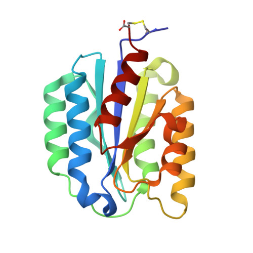
Crystal Structure of the von Willebrand factor A domain of human capillary morphogenesis protein 2: an anthrax toxin receptor
Lacy, D.B., Wigelsworth, D.J., Scobie, H.M., Young, J.A.T., Collier, R.J.(2004) Proc Natl Acad Sci U S A 101: 6367-6372
- PubMed: 15079089 Search on PubMedSearch on PubMed Central
- DOI: https://doi.org/10.1073/pnas.0401506101
- Primary Citation Related Structures:
1SHT, 1SHU - PubMed Abstract:
Anthrax toxin is released from Bacillus anthracis as three monomeric proteins, which assemble into toxic complexes at the surface of receptor-bearing host cells. One of the proteins, protective antigen (PA), binds to receptors and orchestrates the delivery of the other two (the lethal and edema factors) into the cytosol. PA has been shown to bind to two cellular receptors: anthrax toxin receptor/tumor endothelial marker 8 and capillary morphogenesis protein 2 (CMG2). Both are type 1 membrane proteins that include an approximately 200-aa extracellular von Willebrand factor A (VWA) domain with a metal ion-dependent adhesion site (MIDAS) motif. The anthrax toxin receptor/tumor endothelial marker 8 and CMG2 VWA domains share approximately 60% amino acid identity and bind PA directly in a metal-dependent manner. Here, we report the crystal structure of the CMG2 VWA domain, with and without its intramolecular disulfide bond, to 1.5 and 1.8 A, respectively. Both structures contain a carboxylate ligand-mimetic bound at the MIDAS and appear as open conformations when compared with the VWA domains from alpha-integrins. The CMG2 structures provide a template to begin probing the high-affinity CMG2-PA interaction (200 pM) and may facilitate understanding of toxin assembly/internalization and the development of new anthrax treatments. The structural data also allow molecular interpretation of known CMG2 VWA domain mutations linked to the genetic disorders, juvenile hyaline fibromatosis, and infantile systemic hyalinosis.
- Department of Microbiology and Molecular Genetics, Harvard Medical School, 200 Longwood Avenue, Boston, MA 02115, USA.
Organizational Affiliation: