1SE3 | pdb_00001se3

STAPHYLOCOCCAL ENTEROTOXIN B COMPLEXED WITH GM3 TRISACCHARIDE


Experimental Data Snapshot

  • Method: X-RAY DIFFRACTION
  • Resolution: 2.30 Å
  • R-Value Free: 
    0.330 (Depositor) 
  • R-Value Work: 
    0.220 (Depositor) 
  • R-Value Observed: 
    0.220 (Depositor) 

wwPDB Validation 3D Report Full Report

Validation slider image for 1SE3

This is version 2.1 of the entry. See complete history

Literature

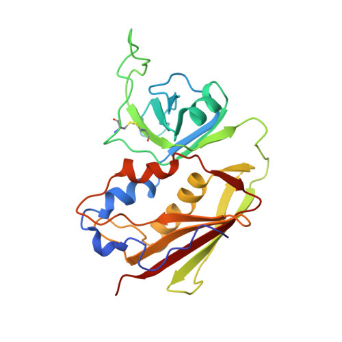
Residues defining V beta specificity in staphylococcal enterotoxins.

Swaminathan, S.Furey, W.Pletcher, J.Sax, M.

(1995) Nat Struct Biol 2: 680-686

  • DOI: https://doi.org/10.1038/nsb0895-680
  • Primary Citation Related Structures: 
    1SE2, 1SE3, 1SE4

  • PubMed Abstract: 

    The three-dimensional structure of staphylococcal enterotoxin C2 has been determined at 2.7 A resolution by x-ray diffraction, while the structures of enterotoxins A and E have been modelled based on their sequence homology to other staphylococcal enterotoxins. The T-cell receptor-binding sites of staphylococcal enterotoxin (SE) B and SEC2 are compared and the stereochemical interactions likely to be responsible for their differing V beta specificities are identified. A similar comparison is made between SEA and SEE.


  • Organizational Affiliation
    • Biocrystallography Laboratory, VA Medical Center, Pittsburgh, Pennsylvania 15240, USA.

Macromolecule Content 

  • Total Structure Weight: 29.04 kDa 
  • Atom Count: 2,153 
  • Modeled Residue Count: 239 
  • Deposited Residue Count: 239 
  • Unique protein chains: 1

Macromolecules

Find similar proteins by:|  3D Structure
Entity ID: 1
MoleculeChains  Sequence LengthOrganismDetailsImage
STAPHYLOCOCCAL ENTEROTOXIN B239Staphylococcus aureusMutation(s): 0 
UniProt
Find proteins for P01552 (Staphylococcus aureus)
Explore P01552 
Go to UniProtKB:  P01552
Entity Groups
Sequence Clusters30% Identity50% Identity70% Identity90% Identity95% Identity100% Identity
UniProt GroupP01552
Sequence Annotations
Expand
Reference Sequence

Oligosaccharides

Help  
Entity ID: 2
MoleculeChains Length2D Diagram GlycosylationD Interactions
N-acetyl-alpha-neuraminic acid-(2-3)-beta-D-galactopyranose-(1-4)-beta-D-glucopyranose
B
3N/A
Glycosylation Resources
GlyTouCan: G91237TK
GlyCosmos: G91237TK
GlyGen: G91237TK

Experimental Data & Validation

Experimental Data

  • Method: X-RAY DIFFRACTION
  • Resolution: 2.30 Å
  • R-Value Free:  0.330 (Depositor) 
  • R-Value Work:  0.220 (Depositor) 
  • R-Value Observed: 0.220 (Depositor) 
Space Group: P 21 21 21
Unit Cell:
Length ( Å )Angle ( ˚ )
a = 45.3α = 90
b = 71β = 90
c = 78.4γ = 90
Software Package:
Software NamePurpose
XENGENdata collection
FBSCALEdata reduction
X-PLORmodel building
X-PLORrefinement
XENGENdata reduction
FBSCALEdata scaling
X-PLORphasing

Structure Validation

View Full Validation Report


Entry History 

Deposition Data

Revision History  (Full details and data files)

  • Version 1.0: 1997-06-16
    Type: Initial release
  • Version 1.1: 2008-03-24
    Changes: Version format compliance
  • Version 1.2: 2011-07-13
    Changes: Version format compliance
  • Version 2.0: 2020-07-29
    Type: Remediation
    Reason: Carbohydrate remediation
    Changes: Atomic model, Data collection, Derived calculations, Other, Structure summary
  • Version 2.1: 2024-10-30
    Changes: Data collection, Database references, Structure summary