1SDS | pdb_00001sds

Structure of protein L7Ae bound to a K-turn derived from an archaeal box H/ACA sRNA


Experimental Data Snapshot

  • Method: X-RAY DIFFRACTION
  • Resolution: 1.80 Å
  • R-Value Free: 
    0.225 (Depositor), 0.232 (DCC) 
  • R-Value Work: 
    0.207 (Depositor), 0.214 (DCC) 
  • R-Value Observed: 
    0.207 (Depositor) 

wwPDB Validation 3D Report Full Report

Validation slider image for 1SDS

This is version 1.5 of the entry. See complete history

Literature

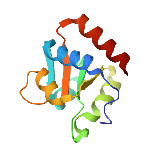
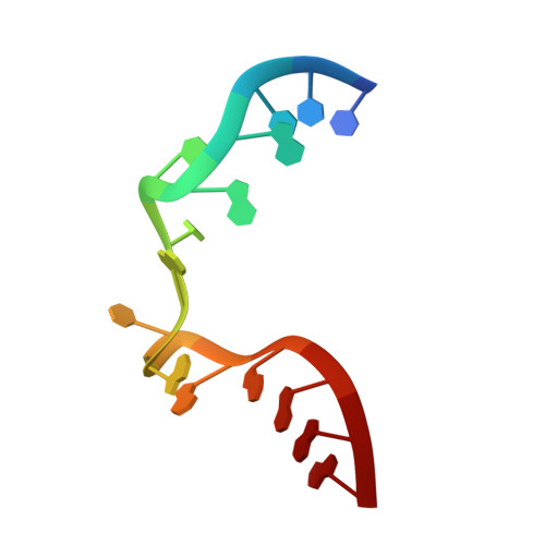
Structure of Protein L7Ae Bound to a K-Turn Derived from an Archaeal Box H/ACA sRNA at 1.8 A Resolution.

Hamma, T.Ferre-D'Amare, A.

(2004) Structure 12: 893-903

  • DOI: https://doi.org/10.1016/j.str.2004.03.015
  • Primary Citation Related Structures: 
    1SDS

  • PubMed Abstract: 

    The archaeal RNA binding protein L7Ae and its eukaryotic homolog 15.5 kDa/Snu13 recognize K-turns. This structural motif is canonically comprised of two stems (one with tandem A.G base pairs, the other with Watson-Crick pairs) linked by an asymmetric internal loop. L7Ae recognizes conventional K-turns in ribosomal and box C/D RNAs but also binds specifically to some box H/ACA RNAs at terminal stem loops. These have the A.G paired stem, but lack the Watson-Crick stem. The structure of Methanococcus jannaschii L7Ae bound to a symmetric duplex RNA without Watson-Crick stems demonstrates how a binding site for this component of diverse ribonucleoprotein complexes can be constructed with only the A.G stem and the loop. The RNA adopts a functional conformation with the aid of a base triple and tight binding of divalent cations. Comparison with the 15.5 kDa/Snu13-RNA complex structure suggests why the eukaryotic homolog does not recognize terminal stem loop L7Ae binding sites.


  • Organizational Affiliation
    • Division of Basic Sciences, Fred Hutchinson Cancer Research Center, 1100 Fairview Avenue North, Seattle, WA 98109 USA.

Macromolecule Content 

  • Total Structure Weight: 53.15 kDa 
  • Atom Count: 3,695 
  • Modeled Residue Count: 381 
  • Deposited Residue Count: 396 
  • Unique protein chains: 1
  • Unique nucleic acid chains: 1

Macromolecules


Find similar proteins by:|  3D Structure
Entity ID: 2
MoleculeChains  Sequence LengthOrganismDetailsImage
50S ribosomal protein L7AeD [auth A],
E [auth B],
F [auth C]
117Methanocaldococcus jannaschiiMutation(s): 0 
Gene Names: RPL7AEMJ1203
UniProt
Find proteins for P54066 (Methanocaldococcus jannaschii (strain ATCC 43067 / DSM 2661 / JAL-1 / JCM 10045 / NBRC 100440))
Explore P54066 
Go to UniProtKB:  P54066
Entity Groups
Sequence Clusters30% Identity50% Identity70% Identity90% Identity95% Identity100% Identity
UniProt GroupP54066
Sequence Annotations
Expand
Reference Sequence
Find similar nucleic acids by:  Sequence
Entity ID: 1
MoleculeChains LengthOrganismImage
box H/ACA sRNAA [auth D],
B [auth E],
C [auth F]
15N/A
Sequence Annotations
Expand
Reference Sequence

Small Molecules

Ligands 2 Unique
IDChains Name / Formula / InChI Key2D Diagram3D Interactions
CA

Query on CA


Download:Ideal Coordinates CCD File
G [auth D]
H [auth D]
I [auth D]
J [auth E]
K [auth E]
G [auth D],
H [auth D],
I [auth D],
J [auth E],
K [auth E],
L [auth E],
N [auth F],
O [auth F],
P [auth F],
Q [auth F]
CALCIUM ION
Ca
BHPQYMZQTOCNFJ-UHFFFAOYSA-N
K

Query on K


Download:Ideal Coordinates CCD File
M [auth E]POTASSIUM ION
K
NPYPAHLBTDXSSS-UHFFFAOYSA-N

Experimental Data & Validation

Experimental Data

  • Method: X-RAY DIFFRACTION
  • Resolution: 1.80 Å
  • R-Value Free:  0.225 (Depositor), 0.232 (DCC) 
  • R-Value Work:  0.207 (Depositor), 0.214 (DCC) 
  • R-Value Observed: 0.207 (Depositor) 
Space Group: F 2 2 2
Unit Cell:
Length ( Å )Angle ( ˚ )
a = 106.97α = 90
b = 141.3β = 90
c = 162.87γ = 90
Software Package:
Software NamePurpose
CNSrefinement
HKL-2000data reduction
SCALEPACKdata scaling
SOLVEphasing

Structure Validation

View Full Validation Report


Entry History 

Deposition Data

Revision History  (Full details and data files)

  • Version 1.0: 2004-05-18
    Type: Initial release
  • Version 1.1: 2008-04-29
    Changes: Version format compliance
  • Version 1.2: 2011-07-13
    Changes: Version format compliance
  • Version 1.3: 2011-11-16
    Changes: Atomic model
  • Version 1.4: 2018-01-31
    Changes: Experimental preparation
  • Version 1.5: 2024-02-14
    Changes: Data collection, Database references, Derived calculations