1SBZ | pdb_00001sbz

Crystal Structure of dodecameric FMN-dependent Ubix-like Decarboxylase from Escherichia coli O157:H7


Experimental Data Snapshot

  • Method: X-RAY DIFFRACTION
  • Resolution: 2.00 Å
  • R-Value Free: 
    0.216 (Depositor), 0.216 (DCC) 
  • R-Value Work: 
    0.176 (Depositor), 0.178 (DCC) 
  • R-Value Observed: 
    0.180 (Depositor) 

wwPDB Validation 3D Report Full Report

Validation slider image for 1SBZ

Ligand Structure Quality Assessment 


This is version 1.5 of the entry. See complete history

Literature

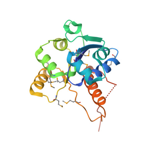
Crystal structure of a dodecameric FMN-dependent UbiX-like decarboxylase (Pad1) from Escherichia coli O157: H7.

Rangarajan, E.S.Li, Y.Iannuzzi, P.Tocilj, A.Hung, L.-W.Matte, A.Cygler, M.

(2004) Protein Sci 13: 3006-3016

  • DOI: https://doi.org/10.1110/ps.04953004
  • Primary Citation Related Structures: 
    1SBZ

  • PubMed Abstract: 

    The crystal structure of the flavoprotein Pad1 from Escherichia coli O157:H7 complexed with the cofactor FMN has been determined by the multiple anomalous diffraction method and refined at 2.0 A resolution. This protein is a paralog of UbiX (3-octaprenyl-4-hydroxybenzoate carboxylyase, 51% sequence identity) that catalyzes the third step in ubiquinone biosynthesis and to Saccharomyces cerevisiae Pad1 (54% identity), an enzyme that confers resistance to the antimicrobial compounds phenylacrylic acids through decarboxylation of these compounds. Each Pad1 monomer consists of a typical Rossmann fold containing a non-covalently bound molecule of FMN. The fold of Pad1 is similar to MrsD, an enzyme associated with lantibiotic synthesis; EpiD, a peptidyl-cysteine decarboxylase; and AtHAL3a, the enzyme, which decarboxylates 4'-phosphopantothenoylcysteine to 4'-phosphopantetheine during coenzyme A biosynthesis, all with a similar location of the FMN binding site at the interface between two monomers, yet each having little sequence similarity to one another. All of these proteins associate into oligomers, with a trimer forming the common structural unit in each case. In MrsD and EpiD, which belong to the homo-dodecameric flavin-containing cysteine decarboxylase (HFCD) family, these trimers associate further into dodecamers. Pad1 also forms dodecamers, although the association of the trimers is completely different, resulting in exposure of a different side of the trimer unit to the solvent. This exposure affects the location of the substrate binding site and, specifically, its access to the FMN cofactor. Therefore, Pad1 forms a separate family, distinguishable from the HFCD family.


  • Organizational Affiliation
    • Biotechnology Research Institute, NRCC, 6100 Royalmount Avenue, Montreal, Quebec H4P 2R2, Canada.

Macromolecule Content 

  • Total Structure Weight: 89.87 kDa 
  • Atom Count: 6,008 
  • Modeled Residue Count: 720 
  • Deposited Residue Count: 788 
  • Unique protein chains: 1

Macromolecules

Find similar proteins by:|  3D Structure
Entity ID: 1
MoleculeChains  Sequence LengthOrganismDetailsImage
Probable aromatic acid decarboxylase
A, B, C, D
197Escherichia coli O157:H7Mutation(s): 11 
Gene Names: PAD1Z4047ECS3593
EC: 4.1.1 (PDB Primary Data), 2.5.1.129 (UniProt)
UniProt
Find proteins for P69772 (Escherichia coli O157:H7)
Explore P69772 
Go to UniProtKB:  P69772
Entity Groups
Sequence Clusters30% Identity50% Identity70% Identity90% Identity95% Identity100% Identity
UniProt GroupP69772
Sequence Annotations
Expand
Reference Sequence

Experimental Data & Validation

Experimental Data

  • Method: X-RAY DIFFRACTION
  • Resolution: 2.00 Å
  • R-Value Free:  0.216 (Depositor), 0.216 (DCC) 
  • R-Value Work:  0.176 (Depositor), 0.178 (DCC) 
  • R-Value Observed: 0.180 (Depositor) 
Space Group: H 3
Unit Cell:
Length ( Å )Angle ( ˚ )
a = 95.372α = 90
b = 95.372β = 90
c = 217.538γ = 120
Software Package:
Software NamePurpose
REFMACrefinement
HKL-2000data reduction
SCALEPACKdata scaling
SOLVEphasing
CNSrefinement

Structure Validation

View Full Validation Report


Ligand Structure Quality Assessment 


Entry History 

Revision History  (Full details and data files)

  • Version 1.0: 2004-10-26
    Type: Initial release
  • Version 1.1: 2008-04-29
    Changes: Version format compliance
  • Version 1.2: 2011-07-13
    Changes: Derived calculations, Source and taxonomy, Version format compliance
  • Version 1.3: 2018-01-31
    Changes: Experimental preparation
  • Version 1.4: 2019-07-24
    Changes: Data collection, Derived calculations, Refinement description
  • Version 1.5: 2024-11-06
    Changes: Data collection, Database references, Derived calculations, Structure summary