1SAT | pdb_00001sat

CRYSTAL STRUCTURE OF THE 50 KDA METALLO PROTEASE FROM S. MARCESCENS


Experimental Data Snapshot

  • Method: X-RAY DIFFRACTION
  • Resolution: 1.75 Å
  • R-Value Work: 
    0.188 (Depositor) 
  • R-Value Observed: 
    0.188 (Depositor) 

wwPDB Validation 3D Report Full Report

Validation slider image for 1SAT

This is version 1.3 of the entry. See complete history

Literature

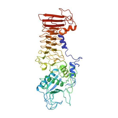
Crystal structure of the 50 kDa metallo protease from Serratia marcescens.

Baumann, U.

(1994) J Mol Biology 242: 244-251

  • DOI: https://doi.org/10.1006/jmbi.1994.1576
  • Primary Citation Related Structures: 
    1SAT

  • PubMed Abstract: 

    The crystal structure of the 50 kDa metalloprotease from the Gram-negative bacterium Serratia marcescens has been solved and refined to a crystallographic R-factor of 0.192 at 1.80 A resolution. The structure is very similar to that of alkaline protease from Pseudomonas aeruginosa, in particular the calcium binding "parallel beta roll" motif is completely conserved. The N-terminal proteolytic domain shows the typical "metzincin" fold. The active sites of the two enzymes are slightly different, Tyr216 is a Zn ligand in the Serratia metallo protease. The loops 70-77 and 122-132, which encompass the active site cleft, differ due to insertions and deletions so that the Serratia metallo protease seems to have a more open site than the alkaline protease.


  • Organizational Affiliation
    • Abteilung für Strukturforschung, Max-Planck-Institut für Biochemie, Martinsried, Germany.

Macromolecule Content 

  • Total Structure Weight: 50.66 kDa 
  • Atom Count: 3,741 
  • Modeled Residue Count: 468 
  • Deposited Residue Count: 471 
  • Unique protein chains: 1

Macromolecules

Find similar proteins by:|  3D Structure
Entity ID: 1
MoleculeChains  Sequence LengthOrganismDetailsImage
SERRATIA PROTEASE471Serratia marcescensMutation(s): 0 
EC: 3.4.24.40
UniProt
Find proteins for P23694 (Serratia marcescens)
Explore P23694 
Go to UniProtKB:  P23694
Entity Groups
Sequence Clusters30% Identity50% Identity70% Identity90% Identity95% Identity100% Identity
UniProt GroupP23694
Sequence Annotations
Expand
Reference Sequence

Experimental Data & Validation

Experimental Data

  • Method: X-RAY DIFFRACTION
  • Resolution: 1.75 Å
  • R-Value Work:  0.188 (Depositor) 
  • R-Value Observed: 0.188 (Depositor) 
Space Group: P 21 21 21
Unit Cell:
Length ( Å )Angle ( ˚ )
a = 151α = 90
b = 109.2β = 90
c = 42.6γ = 90
Software Package:
Software NamePurpose
X-PLORmodel building
X-PLORrefinement
X-PLORphasing

Structure Validation

View Full Validation Report


Entry History 

Deposition Data

  • Released Date: 1995-07-31 
  • Deposition Author(s): Baumann, U.

Revision History  (Full details and data files)

  • Version 1.0: 1995-07-31
    Type: Initial release
  • Version 1.1: 2008-03-24
    Changes: Version format compliance
  • Version 1.2: 2011-07-13
    Changes: Version format compliance
  • Version 1.3: 2024-02-14
    Changes: Data collection, Database references, Derived calculations, Other